Review
. 2010 Apr 15;12(8):1009-59.
doi: 10.1089/ars.2009.2597.

Generation and biological activities of oxidized phospholipids

Affiliations
Review

Generation and biological activities of oxidized phospholipids

Valery N Bochkov et al. Antioxid Redox Signal. .

Abstract

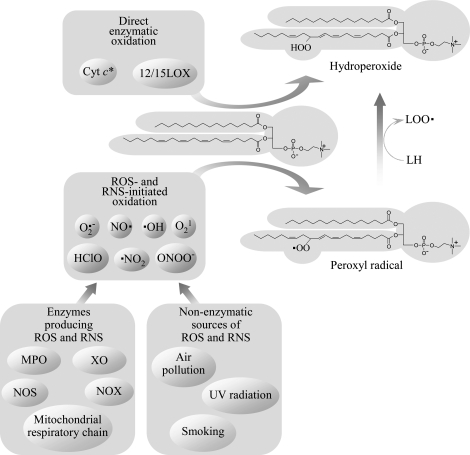
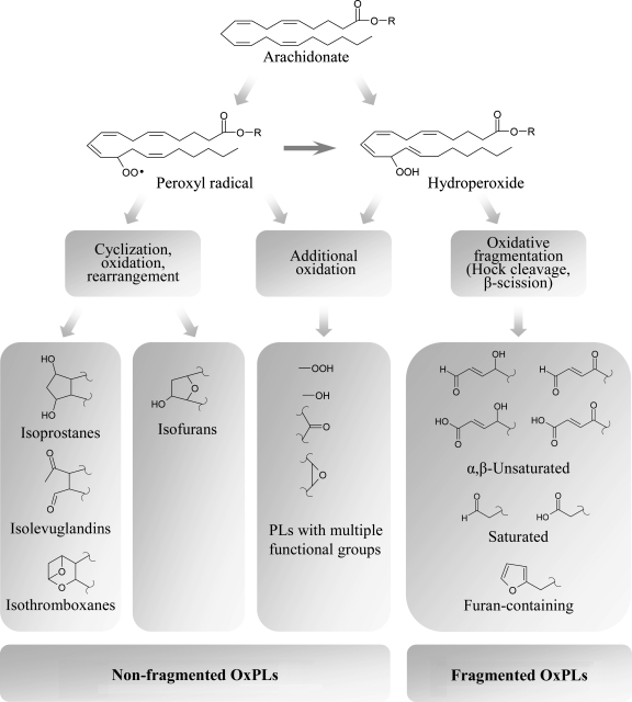
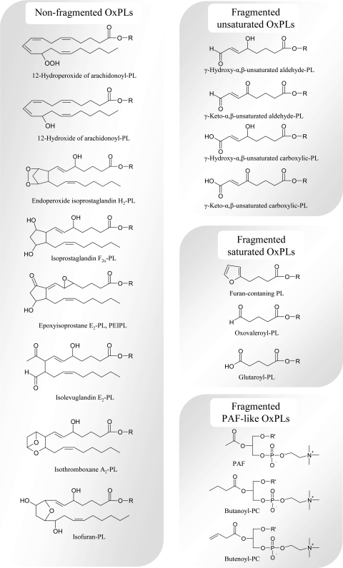
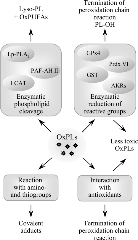
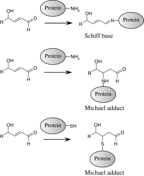
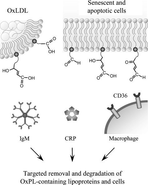
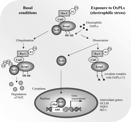
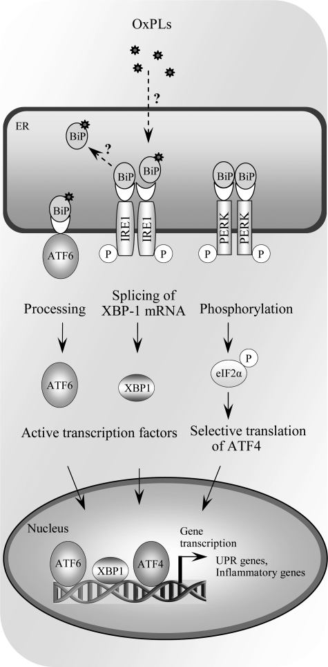
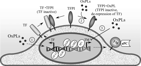
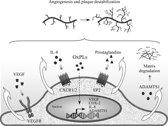
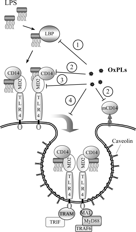
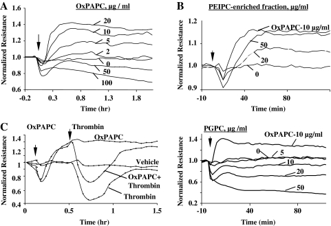
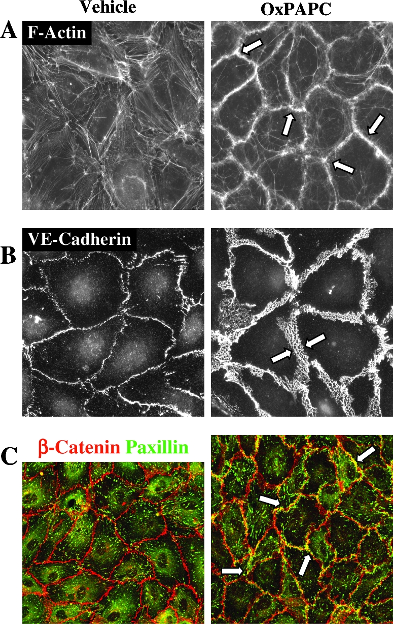
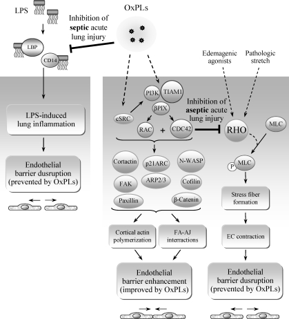
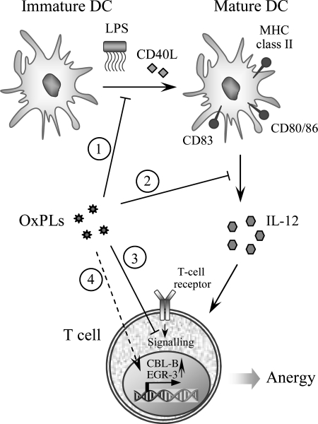
Glycerophospholipids represent a common class of lipids critically important for integrity of cellular membranes. Oxidation of esterified unsaturated fatty acids dramatically changes biological activities of phospholipids. Apart from impairment of their structural function, oxidation makes oxidized phospholipids (OxPLs) markers of "modified-self" type that are recognized by soluble and cell-associated receptors of innate immunity, including scavenger receptors, natural (germ line-encoded) antibodies, and C-reactive protein, thus directing removal of senescent and apoptotic cells or oxidized lipoproteins. In addition, OxPLs acquire novel biological activities not characteristic of their unoxidized precursors, including the ability to regulate innate and adaptive immune responses. Effects of OxPLs described in vitro and in vivo suggest their potential relevance in different pathologies, including atherosclerosis, acute inflammation, lung injury, and many other conditions. This review summarizes current knowledge on the mechanisms of formation, structures, and biological activities of OxPLs. Furthermore, potential applications of OxPLs as disease biomarkers, as well as experimental therapies targeting OxPLs, are described, providing a broad overview of an emerging class of lipid mediators.


Figures

FIG. 1.
FIG. 1.
Mechanisms initiating peroxidation of PUFAs esterified in phospholipids. Peroxidation of phospholipids containing PUFAs is initiated via both enzymatic and nonenzymatic mechanisms. Lipoxygenases from the 12/15 family accept PL-esterified PUFAs as substrate and insert dioxygen, producing hydroperoxides. PUFA peroxidation can also be induced by nonradical ROS (singlet oxygen), or by free radicals, which either penetrate from the environment, or are produced endogenously by enzymes, such as NADPH oxidase (NOX), myeloperoxidase (MPO), nitric oxide synthase (NOS), xanthine oxidase (XO), or respiratory chain in mitochondria. Reactions induced by different free radicals followed by addition of oxygen produce the same type of primary oxidation products (i.e., peroxyl radicals), which in turn transform into hydroperoxides after reacting with other PUFA molecules. *Cyt c is selective for cardiolipin and phosphatidylserine as compared to PC (164).
FIG. 2.
FIG. 2.
Oxidation of esterified PUFA generates a variety of nonfragmented and fragmented OxPLs. Top panel presents mass spectrum of synthetic palmitoyl-arachidonoyl-phosphatidylcholine (PAPC). Bottom panel shows multiple products generated from PAPC upon prolonged exposure of pure dry lipid to air. Note formation of multiple fragmented (m/z < 782) and nonfragmented (m/z > 782) OxPAPC species that were generated by nonenzymatic oxidation of just one precursor molecule.
FIG. 3.
FIG. 3.
Evolution of phospholipid oxidation products. Peroxidation of PL-esterified PUFAs is initiated by formation of hydroperoxides or peroxyl radicals. Further evolution of primary PL oxidation products proceeds without participation of enzymes via three major pathways. First, additional oxidation within the same PUFA generates OxPLs with various combinations of functional groups such as hydroperoxides, hydroxides, keto- and epoxy-groups. Second pathway involves intramolecular cyclization, rearrangement, and further oxidation. If bicyclic endoperoxide is formed as an intermediate product, three groups of products are generated, including isoprostanes, isolevuglandins, and isothromboxanes, while cyclization leading to formation of monocyclic peroxide finally produces isofurans. Third group of transformations results from several chemical reactions all leading to fragmentation of PUFAs and generation of short residues having various combinations of hydroxide and carbonyl groups, or terminal furan.
FIG. 4.
FIG. 4.
Major forms of oxidatively modified residues that were identified in OxPLs. The figure presents example structures from all groups of PUFA oxidation products that were shown to exist in PL-esterified form. R is 1-acyl-2-lyso-sn-glycero-3-X; X, phosphocholine, phosphoserine, phosphoethanolamine, phosphoinositol, phosphoglycerol, phosphate; R' is an alkyl residiue.
FIG. 5.
FIG. 5.
Termination of phospholipid oxidation. Several processes play a role in termination of peroxidation chain reaction and detoxification of reactive groups in PL-esterified PUFAs. In addition to scavenging of radicals by antioxidants, reactive peroxide groups are reduced by specific form of glutathione peroxidase (GPx4) capable of reducing PL-esterified residues, as well as peroxiredoxin VI and glutathione transferase (GST). Reactive carbonyl groups in PL residues are reduced by aldo-keto-reductases from AKR1A and B families. Furthermore, several phospholipases A selectively cleave oxidized residues, leading to formation of lyso-PLs and free oxidized fatty acids. Similar activity is demonstrated by LCAT. Finally, electrophilic PLs can form covalent complexes with amino acids, which may inactivate reactive groups on PLs but on the other hand can damage sensitive proteins.
FIG. 6.
FIG. 6.
Chemical reactions of OxPLs containing ϖ-terminal aldehyde groups with amino- and sulfhydryl groups in proteins. OxPL residues having reactive carbonyl groups can form Schiff bases or Michael adducts with NH2- and SH-groups of proteins. Formation of covalent complexes can modulate activity and half-life of OxPLs, initiate electrophilic stress response and inactivate sensitive proteins.
FIG. 7.
FIG. 7.
OxPLs are recognized by soluble and cell-associated pattern-recognition receptors of innate immunity. Fragmented oxidized residues change their orientation within the cell membrane or lipoprotein outer layer, and protrude into the water phase, thus enabling recognition by cellular receptors (e.g., CD36, natural (germ-line encoded) immunoglobulins or C-reactive protein). It is likely that in analogy with other, better characterized ligands of these proteins, interactions with OxPLs result in removal and degradation of oxidized lipoproteins, senescent and apoptotic cells. Furthermore, binding of OxPLs to CD36 promotes formation of foam cells characteristic of atherosclerosis.
FIG. 8.
FIG. 8.
Activation of electrophilic stress response by OxPLs. Induction of antioxidant genes is initiated by reactive electrophilic compounds via covalent modification of thiol groups in KEAP1 leading to derepression of transcription factor NRF2. OxPLs were shown to induce nuclear accumulation of NRF2, stimulate its binding to promoters of target genes, and elevate mRNA and protein levels of major antioxidant genes. It has to be established whether OxPLs similarly to classical electrophiles form covalent complexes with thiol groups of KEAP1.
FIG. 9.
FIG. 9.
Activation of unfolded protein response (UPR) by OxPLs. Multiple stress conditions leading to impaired processing of proteins in endoplasmic reticulum activate adaptive reaction called UPR, which is mediated via three branches, all initiated by binding of unfolded proteins to BIP/GRP78, thus leading to dissociation from, and de-repression of, ATF6, IRE1, and PERK. Dissociation of BIP/GRP78 initiates processing of ATF6 protein, splicing of XBP-1 mRNA by IRE1, and phosphorylation of eIF2α, resulting in selective translation of ATF4. OxPAPC and other classes of OxPLs were shown to activate all three arms of the UPR, leading to formation of transcriptionally active ATF6, XBP1, and ATF4, as well as induction of their target genes. However, the mechanism of activation is currently unknown. In particular, it is not clear whether similarly to unfolded proteins OxPLs directly bind to BiP/GRP78 and reverse repression of ATF6, IRE1, and PERK.
FIG. 10.
FIG. 10.
OxPLs induce pro-coagulant shift in endothelium. OxPLs were shown to activate three pro-coagulant mechanisms in endothelium. First, OxPAPCs was shown to upregulate via EGR-1- and NFAT-dependent mechanisms expression and activity of the major inducer of coagulation, tissue factor (TF, mechanism 1). Furthermore, peroxidation products of PC and PE containing oxidized linoleic acid were shown to inactivate tissue factor pathway inhibitor (TFPI, mechanism 2) as a result of direct binding of OxPLs to the C-terminus of TFPI. Finally, OxPAPC was shown to inhibit expression of the major anticoagulant protein on ECs, thrombomodulin (TM, mechanism 3). The effect was explained by decreased activity of transcription factors mediating basal expression of TM (i.e., retinoic acid receptor β, retinoid X receptor α, Sp1 and Sp3).
FIG. 11.
FIG. 11.
OxPLs induce angiogenic shift in ECs via autocrine mechanisms. OxPAPC and other classes of oxidized diacyl-OxPLs, but not their unoxidized precursors, stimulate angiogenic reactions in several in vitro and in vivo models. Angiogenic effects of OxPLs are mediated by autocrine loops, including production of VEGF, IL-8, and COX2-derived prostaglandins. In parallel, OxPLs stimulate expression of metalloproteinase ADAMTS1. Together, these effects are likely to promote formation of neovessels and degradation of matrix, thus leading to destabilization of atherosclerotic plaque.
FIG. 12.
FIG. 12.
Potential mechanisms of the anti-endotoxin action of OxPLs. Different classes and molecular species of OxPLs were shown to inhibit effects of LPS in vitro and in vivo. OxPLs were hypothesized to inhibit several steps in recognition of LPS by TLR4 and activation of downstream signaling events. OxPAPC and other classes of OxPLs were shown to bind to LBP, soluble and membrane-bound CD14, and MD-2, and thus to inhibit interactions of these proteins with LPS, which are critically important for activation of TLR4 (mechanisms 1–3). In addition, OxPLs were shown to disrupt lipid rafts thus preventing formation of signaling complex of TLR4 with intracellular adaptors within caveolin-rich membrane domains (mechanism 4). Thus, OxPLs inhibit action of bacterial endotoxin via a multi-hit mechanism.
FIG. 13.
FIG. 13.
Effects of oxidized phospholipids on endothelial barrier function. (A) Bi-phasic concentration-dependent effects of OxPAPC on endothelial barrier function. Transendothelial electrical resistance (TER) reflecting barrier properties of EC monolayer was recorded in pulmonary ECs exposed to OxPAPC. OxPAPC exhibited prominent barrier-protective effects at concentrations below 20 μg/ml. Higher OxPAPC concentrations (50 and 100 μg/ml) caused barrier-disruptive response. (B) Nonfragmented, but not oxidatively fragmented PLs exhibit barrier-protective effect. TER was measured in pulmonary EC monolayers exposed to HPLC-purified isoprostanes esterified in PC (upper panel), or fragmented product PGPC (lower panel). Another fragmented product POVPC exhibited disruptive effects similar to that shown for PGPC. Note that individual components of OxPAPC, such as PEIPC and PGPC, demonstrate opposite action on EC barrier. Adapted from (28). (C) OxPAPC attenuates thrombin-induced elevation of EC permeability. TER was monitored across the confluent EC monolayers treated with OxPAPC (20 μg/ml) and thrombin (0.5 U/ml) added at the times shown by arrows. Adapted from (31).
FIG. 14.
FIG. 14.
OxPAPC-induced endothelial remodeling. Enhancement of peripheral endothelial actin cytoskeleton (A, arrows), VE-cadherin positive adherens junctions (B, arrows), and peripheral colocalization of focal adhesions and adherens junctions (C, arrows) detected by double immunofluorescent staining for β-catenin (red) and paxillin (green) and confocal microscopy. ECs were stimulated with barrier-protective OxPAPC concentration (20 μg/ml). Adapted from (31, 33).
FIG. 15.
FIG. 15.
Protective effects of OxPLs against acute lung injury and endothelial barrier dysfunction. PL oxidation products such as OxPAPC can inhibit lung damage in acute bacterial inflammation due to their ability to inhibit activation of TLRs 2 and 4. Furthermore, PL-esterified isoprostanes activate tyrosine kinase SRC and PI3K-AKT signaling, leading to recruitment of RAC/CDC42 specific nucleotide exchange factors TIAM1 and βPIX, stimulation of RAC and CDC42 GTPases and their effectors involved in activation of peripheral actin polymerization (cortactin, p21ARC, ARP2/3, N-WASP, cofilin), focal adhesion remodeling (FAK, paxillin) and enhancement of adherens junctions (β-catenin). These cytoskeletal changes are critical for enhancement of vascular endothelial barrier properties. Finally, OxPAPC and other classes of OxPLs protect against vascular hyperpermeability caused by edemagenic, inflammatory factors, and pathologic lung overdistension. This protection involves inhibition of RHO GTPase-dependent pathway of endothelial contraction and monolayer barrier disruption via activation of RAC-dependent mechanisms triggered by OxPLs.
FIG. 16.
FIG. 16.
Inhibitory effects of OxPLs in adaptive immunity. OxPLs can influence adaptive immune responses via several mechanisms. First, OxPLs inhibit maturation of dendritic cells (DCs) induced by LPS and CD40L (mechanism 1). Furthermore, OxPLs inhibit production of IL-12 by DCs (mechanism 2). In addition, several classes of OxPLs were shown to inhibit activation of T cell receptor (mechanism 3). Finally, OxPLs via as yet unidentified mechanisms upregulate expression in T cells of transcription factors CBL-B and EGR-3 inducing an anergy-like state (mechanism 4). Together, these effects characterize OxPLs as immunosuppressors.

References

    1. Ago T. Kitazono T. Ooboshi H. Iyama T. Han YH. Takada J. Wakisaka M. Ibayashi S. Utsumi H. Iida M. Nox4 as the major catalytic component of an endothelial NAD(P)H oxidase. Circulation. 2004;109:227–233. - PubMed
    1. Alcaraz MJ. Fernandez P. Guillen MI. Anti-inflammatory actions of the heme oxygenase-1 pathway. Curr Pharm Des. 2003;9:2541–2551. - PubMed
    1. Ameli S. Hultgardh-Nilsson A. Regnstrom J. Calara F. Yano J. Cercek B. Shah PK. Nilsson J. Effect of immunization with homologous LDL and oxidized LDL on early atherosclerosis in hypercholesterolemic rabbits. Arterioscler Thromb Vasc Biol. 1996;16:1074–1079. - PubMed
    1. Androulakis N. Durand H. Ninio E. Tsoukatos DC. Molecular and mechanistic characterization of platelet-activating factor-like bioactivity produced upon LDL oxidation. J Lipid Res. 2005;46:1923–1932. - PubMed
    1. Anton R. Camacho M. Puig L. Vila L. Hepoxilin B3 and its enzymatically formed derivative trioxilin B3 are incorporated into phospholipids in psoriatic lesions. J Invest Dermatol. 2002;118:139–146. - PubMed

Publication types

MeSH terms