Review
. 2009:43:223-49.
doi: 10.1146/annurev-genet-102108-134222.

How the fanconi anemia pathway guards the genome

Affiliations
Review

How the fanconi anemia pathway guards the genome

George-Lucian Moldovan et al. Annu Rev Genet. 2009.

Abstract

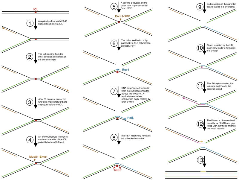
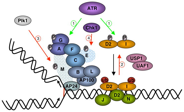
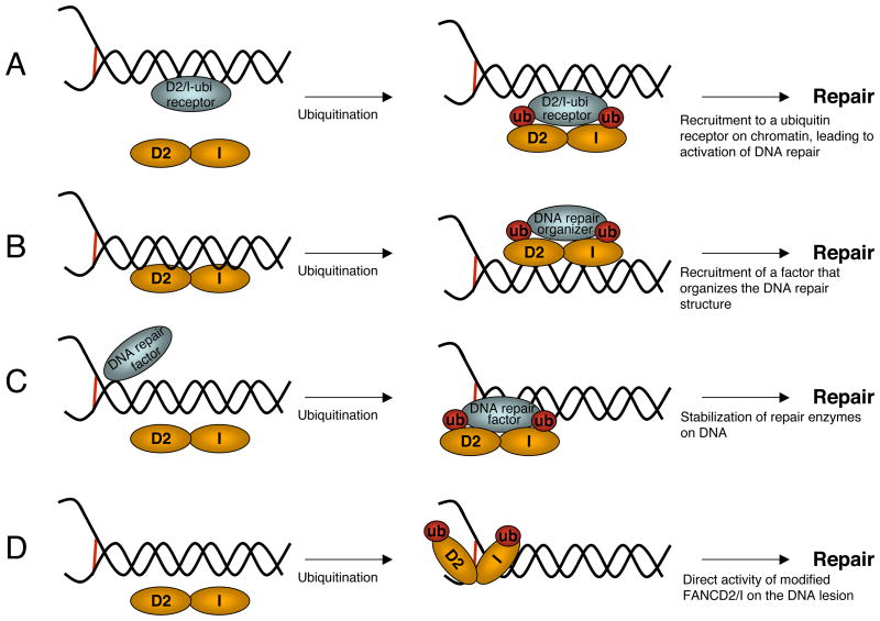
Fanconi Anemia (FA) is an inherited genomic instability disorder, caused by mutations in genes regulating replication-dependent removal of interstrand DNA crosslinks. The Fanconi Anemia pathway is thought to coordinate a complex mechanism that enlists elements of three classic DNA repair pathways, namely homologous recombination, nucleotide excision repair, and mutagenic translesion synthesis, in response to genotoxic insults. To this end, the Fanconi Anemia pathway employs a unique nuclear protein complex that ubiquitinates FANCD2 and FANCI, leading to formation of DNA repair structures. Lack of obvious enzymatic activities among most FA members has made it challenging to unravel its precise modus operandi. Here we review the current understanding of how the Fanconi Anemia pathway components participate in DNA repair and discuss the mechanisms that regulate this pathway to ensure timely, efficient, and correct restoration of chromosomal integrity.


Figures

Figure 1
Figure 1
The Fanconi Anemia pathway protects against genomic instability. (a) Loss of the FA pathway leads to chromosomal aberrations, with a specific increase in the frequency of radial chromosomes following induction of ICLs. Shown is a metaphase spread of a Fanconi Anemia cell. (b) Schematic representation of the FA protein complex: FANCM and FAAP24 recruit a large multisubunit ubiquitin ligase, termed the core complex, to DNA lesions. This structure is composed of subcomplexes (shown in different colors): FANCA-FANCG, FANCC-FANCE-FANCF and FANCB-FANCL-FAAP100. The core complex monoubiquitinates FANCD2 and FANCI, which then localize to DNA repair foci together with FANCD1, FANCJ, and FANCN.
Figure 2
Figure 2
Speculative model for the coordinated use of multiple classic DNA repair pathways in replication-dependent repair of ICLs (adapted from ref. 119). Replication forks converging from different directions stall at an ICL site, 20–40 nucleotides before the lesion (steps 1 and 2). Subsequently, one fork moves forward and stops just before the lesion (step 3). Dual incision on each side of the ICL, likely performed by Mus81-Eme1 (the proximal –step 4) and Ercc1-XPF (the distal –step 5) unhook the ICL, which is then bypassed by a TLS polymerase, probably Rev1 (step 6). DNA polymerase ζ extends from this initial insertion (step 7), and the NER system removes the bypassed crosslink (step 8). The HR machinery then repairs the broken chromatid using the newly repaired sister as a template (steps 9–13). (It is also possible that a double Holliday junction is the relevant structure of the HR reaction –steps 11 and 12.)
Figure 3
Figure 3
Regulation of the FA pathway. Positive regulation is marked by green arrows, and negative control is represented by red arrows. (a) ATR initiates the pathway by phosphorylating FANCI, FANCD2, FANCA, and possibly FANCG. (b) The deubiquitinating enzyme USP1 and its partner and activator UAF1 remove ubiquitin from FANCD2 and FANCI, thus suppressing the pathway. (c) In mitosis, the Plk1 kinase phophorylates FANCM, creating a recognition motif for the ubiquitin ligase βTRCP. FANCM becomes multiubiquitinated and degraded by the proteasome. (d) Chk1-dependent phosphorylation of FANCE leads to its degradation.
Figure 4
Figure 4
Models for the function of FANCD2-I ubiquitination. (a) ubiquitination allows FANCD2-I binding to a nuclear receptor on chromatin, leading to its accumulation in foci and activation of DNA repair. (b) Ubiquitinated FANCD2-I recruit factors that organize the repair response and bind other repair enzymes. (c) Modified FANCD2-I complexes stabilize, activate, or enhance the enzymatic activities of repair factors. (d) Ubiquitination leads to a structural change in FANCD2-I that activates a cryptic DNA repair activity of the complex. These models are not mutually exclusive.

References

    1. Agoulnik AI, Lu B, Zhu Q, Truong C, Ty MT, et al. A novel gene, Pog, is necessary for primordial germ cell proliferation in the mouse and underlies the germ cell deficient mutation, gcd. Hum Mol Genet. 2002;11:3047–53. - PubMed
    1. Akkari YM, Bateman RL, Reifsteck CA, Olson SB, Grompe M. DNA replication is required to elicit cellular responses to psoralen-induced DNA interstrand cross-links. Mol Cell Biol. 2000;20:8283–89. - PMC - PubMed
    1. Alpi A, Langevin F, Mosedale G, Machida YJ, Dutta A, Patel KJ. UBE2T, the Fanconi anemia core complex, and FANCD2 are recruited independently to chromatin: a basis for the regulation of FANCD2 monoubiquitination. Mol Cell Biol. 2007;27:8421–30. - PMC - PubMed
    1. Alpi AF, Pace PE, Babu MM, Patel KJ. Mechanistic insight into site-restricted monoubiquitination of FANCD2 by Ube2t, FANCL, and FANCI. Mol Cell. 2008;32:767–77. - PubMed
    1. Alter BP. Fanconi’s anemia and malignancies. Am J Hematol. 1996;53:99–110. - PubMed

Publication types

Substances

LinkOut - more resources