. 2009 Oct 12;423(3):389-400.
doi: 10.1042/BJ20090086.

Functional characterization of the interactions between endosomal adaptor protein APPL1 and the NuRD co-repressor complex

Affiliations

Functional characterization of the interactions between endosomal adaptor protein APPL1 and the NuRD co-repressor complex

Magdalena Banach-Orlowska et al. Biochem J. .

Abstract

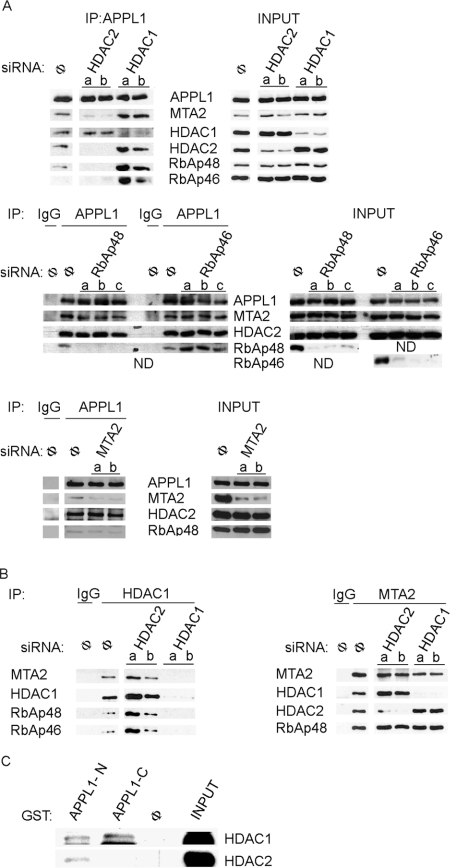
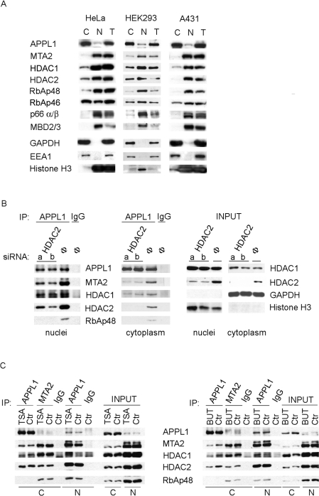
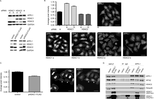
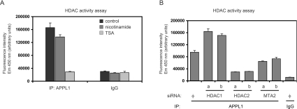
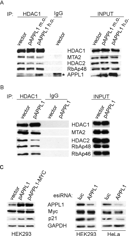
Multifunctional adaptor protein APPL1 [adaptor protein containing PH (pleckstrin homology) domain, PTB (phosphotyrosine binding) domain and leucine zipper motif] belongs to a growing group of endocytic proteins which actively participate in various stages of signalling pathways. Owing to its interaction with the small GTPase Rab5, APPL1 localizes predominantly to a subpopulation of early endosomes but is also capable of nucleocytoplasmic shuttling. Among its various binding partners, APPL1 was reported to associate with the nuclear co-repressor complex NuRD (nucleosome remodelling and deacetylase), containing both nucleosome remodelling and HDAC (histone deacetylase) activities, but the biochemical basis or functional relevance of this interaction remained unknown. Here we characterized the binding between APPL1 and NuRD in more detail, identifying HDAC2 as the key NuRD subunit responsible for this association. APPL1 interacts with the NuRD complex containing enzymatically active HDAC2 but not HDAC1 as the only deacetylase. However, the cellular levels of HDAC1 can regulate the extent of APPL1-NuRD interactions, which in turn modulates the nucleocytoplasmic distribution of APPL1. Increased binding of APPL1 to NuRD upon silencing of HDAC1 promotes the nuclear localization of APPL1, whereas HDAC1 overexpression exerts an opposite effect. Moreover, we also uncovered a NuRD-independent interaction of APPL1 with HDAC1. APPL1 overexpression affects the composition of the HDAC1-containing NuRD complex and the expression of HDAC1 target p21WAF1/CIP1. Cumulatively, these data reveal a surprising complexity of APPL1 interactions with HDACs, with functional consequences for the modulation of gene expression. In a broader sense, these results contribute to an emerging theme of endocytic proteins playing alternative roles in the cell nucleus.


Figures

Figure 1
Figure 1. HDAC2 is critical for binding of APPL1 to the NuRD complex
(A and B) Extracts from HeLa cells transfected for 72 h with two (a, b) or three (a, b, c) different siRNA oligonucleotides per gene against: HDAC1, HDAC2, MTA2, RbAp48 and RbAp46 or non-specific siRNA (Φ) were subjected to immunoprecipitation (IP) using: (A) anti-APPL1 antibody; (B) anti-HDAC1 antibody (left panel) or anti-MTA2 antibody (right panel). Non-specific antibodies (IgG) were used as controls. Input indicates 10% of total cell extracts used for immunoprecipitation. Immunoprecipitates and input extracts were analysed by Western blotting using different antibodies as indicated. (C) To verify the direct interactions between APPL1 and HDAC1 or HDAC2, in vitro translated HDAC1–FLAG and untagged HDAC2 were subjected to GST pull-down assay using GST alone (Φ) or GST fused to the N- or C-terminal parts of APPL1 (APPL1-N or APPL1-C, respectively). Input indicates 10% of in vitro translated material used for the pull-down assay. Bound proteins were analysed by Western blotting using anti-HDAC1 and anti-HDAC2 antibodies. ND, not determined.
Figure 2
Figure 2. APPL1 interacts with the NuRD subunits in both cytoplasmic and nuclear fractions independently of HDAC enzymatic activity
(A) Cytoplasmic (C) and nuclear (N) fractions along with total extracts (T) of three different cell lines, HeLa, HEK-293 and A431, were analysed for the presence of several NuRD subunits by Western blotting with different antibodies as indicated. For detection with a given antibody, equal amounts of proteins from all fractions and three cell lines were loaded (20 μg of protein for blotting with anti-p66α/β, -MBD2/3 and -EEA1 antibodies; 15 μg of protein for anti-APPL1, -HDAC2, -RbAp46 and -GAPDH; 10 μg of protein for anti-MTA2, -HDAC1, -RbAp48 and -Histone H3; the different amounts of protein loaded were chosen to match different sensitivities of the antibodies used). Cytoplasmic (GAPDH and EEA1) and nuclear (histone H3) markers were used to demonstrate the purity of fractions. (B) HeLa cells were transfected for 72 h with two oligonucleotides (a, b) against HDAC2 or with non-specific control oligonucleotide (Φ). Cytoplasmic and nuclear fractions were prepared and subjected to immunoprecipitation (IP) using anti-APPL1 antibody or non-specific immunoglobulins (IgG). Immunoprecipitates were tested for the presence of several NuRD subunits by immunoblotting with various antibodies as indicated. Right panel: 10% of the input material (cytoplasmic and nuclear fractions) were analysed for the knockdown efficiency using anti-HDAC1 and anti-HDAC2 antibodies, as well as for the fraction purity with anti-GAPDH and anti-histone H3 antibodies. (C) Immunoprecipitation from cytoplasmic (C) or nuclear (N) fractions of HeLa cells treated for 20 h with 100 ng/ml of TSA (left panel) or 25 mM sodium butyrate (BUT; right panel) was performed using anti-APPL1, anti-MTA2 or non-specific rabbit IgG. Precipitates along with 10% of the input material were analysed by Western blotting using different antibodies, as indicated. Ctr, control.
Figure 3
Figure 3. The interactions with NuRD affect cellular distribution of APPL1
(A) APPL1 protein levels do not depend on HDAC1 or HDAC2. Extracts of HeLa cells with reduced HDAC1 or HDAC2 levels by siRNA [two different oligonucleotides (a, b) per gene or non-specific oligo (Φ), transfected for 72 h; top panel] and HEK-293 cells transfected for 48 h with plasmids encoding FLAG-tagged HDAC1 (pHDAC1–FLAG), HDAC2 (pHDAC2–FLAG) or with a control vector (bottom panel), were analysed by Western blotting using anti-APPL1 antibodies. To demonstrate the efficiency of silencing or overexpression, extracts were probed using anti-HDAC1 and anti-HDAC2 antibodies. GAPDH was included as a loading control. (B) Microscopy-based analysis of APPL1 nuclear localization upon silencing of HDAC1 or HDAC2. HeLa cells were transfected with siRNA oligonucleotides: one non-specific (Φ) and two specific per gene (a and b) against HDAC1 and HDAC2 for 72 h, followed by fixation and immunostaining for APPL1. Acquired microscopic images were analysed by Metamorph software and the average pixel intensities corresponding to APPL1 in the nuclei (as visualized by Hoechst staining, not shown) were calculated. The results of a representative experiment are shown in the graph. The values are normalized with respect to the average pixel intensity of nuclear APPL1 in cells transfected with non-specific siRNA, assigned one arbitrary unit. Error bars indicate standard error (minimum 100 cells from each transfection were used for the analysis). The results were statistically analysed by GraphPad Prism4 software, and the values obtained for each knockdown experiment were significantly different from the control at P<0.0001. The images demonstrate the cellular localization of APPL1 upon silencing of HDAC1 or HDAC2 (as indicated) and represent a maximal projection of z-stacks. Scale bar: 24 μm. (C) HeLa cells transfected with the plasmid encoding FLAG-tagged HDAC1 (pHDAC1–FLAG) were analysed with respect to the nuclear localization of APPL1 as described above. Untransfected cells (marked with asterisks) were used as a control (the average pixel intensity of nuclear APPL1 set to one unit). The statistical analysis was performed on 50 transfected and 50 untransfected cells, and the difference between them was statistically significant at P<0.0001. The images represent a maximal projection of z-stacks. FLAG staining (left image) is shown to discriminate between transfected and untransfected cells with respect to APPL1 staining (right image). Scale bar: 24 μm. (D) HDAC1 overexpression leads to the destabilization of binding between APPL1 and HDAC2-containing NuRD complex. Extracts of HEK-293 cells transfected with HDAC1-FLAG or with a control vector were subjected to immunoprecipitation (IP) with anti-APPL1 or non-specific (IgG) antibodies. Immunoprecipitates, along with 10% of the extracts used (input), were analysed by Western blotting using the indicated antibodies. MW indicates a lane loaded with a molecular mass (weight) marker. Two exposures (short and long) of the HDAC1 blot are shown. Overexpressed HDAC1 exhibits some non-specific binding to IgG-covered protein G beads (visible at the long exposure of the blot and marked with an asterisk); however, its binding to beads containing anti-APPL1 antibodies is higher.
Figure 4
Figure 4. Deacetylase activity detected in APPL1 complexes derives mainly from HDAC2
HDAC enzymatic activity was measured using a fluorimetric method (as described in the Experimental section) in immunoprecipitates from HeLa cells. (A) APPL1 binds active HDACs from class I or II. The APPL1 immunoprecipitate (IP) was divided into three equal parts: one left untreated and the other two treated with HDAC inhibitors: 1 mM nicotinamide and 1 μM TSA. Non-specific rabbit IgG was used as a control. (B) HeLa cells were silenced for HDAC1, HDAC2 and MTA2 [using two (a, b) different siRNA oligonucleotides per gene or non-specific siRNA Φ] prior to immunoprecipitation using APPL1 antibodies or non-specific rabbit IgG. The same extracts as presented in Figure 1(A) were used (one third of the immunoprecipitates was measured in the HDAC activity assay, two thirds blotted as shown in Figure 1A). The intensity of fluorescence emitted by the deacetylated substrate is expressed in arbitrary units in (A) and (B).
Figure 5
Figure 5. APPL1 overexpression affects the composition of HDAC1-containing NuRD complex and the expression of HDAC1 target p21WAF1/CIP
(A) APPL1 overexpression impairs the interactions of HDAC1 with other NuRD subunits. HEK-293 cells overexpressing untagged APPL1 at moderate or high levels (m.o., moderate overexpression of pAPPL1; h.o., high overexpression of pAPPL1) were subjected to immunoprecipitation (IP) with anti-HDAC1 or non-specific rabbit (IgG) antibodies. Immunoprecipitates along with 10% of the extracts used (input, right panel) were tested by immunoblotting for the presence of several NuRD subunits, as indicated. Some non-specific binding of APPL1 to IgG-covered Protein G beads is marked with an asterisk. (B) APPL1 overexpression reduces the association of HDAC1 with other NuRD components in the nuclear fraction. HDAC1 was immunoprecipitated from the nuclear extracts of HEK-293 cells with endogenous (vector) or overexpressed APPL1. Immunoprecipitates and 5% of the starting material (input, right panel) were blotted for the presence of the indicated NuRD components. (C) APPL1 influences the level of HDAC1 target gene product p21WAF1/CIP1. The level of p21WAF1/CIP1 expression was analysed by Western blotting using anti-p21 antibody in extracts of cells with overexpression or silenced expression of APPL1. Left panel: Extracts of HEK-293 cells overexpressing APPL1 (either untagged, pAPPL1, or MYC-tagged, pAPPL1–MYC) or transfected with a control vector for 48 h were immunoblotted as indicated. No efficient overexpression of APPL1 could be achieved in HeLa cells. Right panel: APPL1 expression was reduced by esiRNA against APPL1 in HEK-293 or HeLa cells, using esiRNA against luciferase (luc) as a specificity control. Transfections with esiRNA were performed for 72 h (HEK-293) or 48 h (HeLa). The resulting extracts were immunoblotted against APPL1, Myc and p21. GAPDH was included as a loading control.

References

    1. Hoeller D., Volarevic S., Dikic I. Compartmentalization of growth factor receptor signalling. Curr. Opin. Cell Biol. 2005;17:107–111. - PubMed
    1. Le Roy C., Wrana J. L. Clathrin- and non-clathrin-mediated endocytic regulation of cell signalling. Nat. Rev. Mol. Cell. Biol. 2005;6:112–126. - PubMed
    1. Miaczynska M., Pelkmans L., Zerial M. Not just a sink: endosomes in control of signal transduction. Curr. Opin. Cell Biol. 2004;16:400–406. - PubMed
    1. Polo S., Di Fiore P. P. Endocytosis conducts the cell signaling orchestra. Cell. 2006;124:897–900. - PubMed
    1. von Zastrow M., Sorkin A. Signaling on the endocytic pathway. Curr. Opin. Cell Biol. 2007;19:436–445. - PMC - PubMed

Publication types

MeSH terms