

Development and regeneration of the inner ear
- PMID: 19686102
- PMCID: PMC7245053
- DOI: 10.1111/j.1749-6632.2009.04484.x
Development and regeneration of the inner ear
Abstract
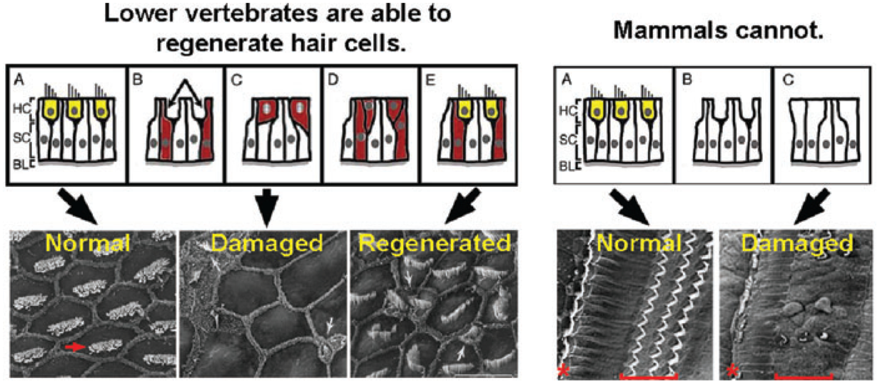
Loss of sensory hair cells is the leading cause of deafness in humans. The mammalian cochlea cannot regenerate its complement of sensory hair cells. Thus at present, the only treatment for deafness due to sensory hair cell loss is the use of prosthetics, such as hearing aids and cochlear implants. In contrast, in nonmammalian vertebrates, such as birds, hair cell regeneration occurs following the death of hair cells and leads to the restoration of hearing. Regeneration in birds is successful because supporting cells that surround the hair cells can divide and are able to subsequently differentiate into new hair cells. However, supporting cells in mammals do not normally divide or transdifferentiate when hair cells are lost, and so regeneration does not occur. To understand the failure of mammalian cochlear hair cell regeneration, we need to understand the molecular mechanisms that underlie cell division control and hair cell differentiation, both during embryogenesis and in the postnatal mouse. In this review, we present a discussion of the regulation of cell proliferation in embryogenesis and during postnatal maturation. We also discuss the role of the Cip/Kip cell cycle inhibitors and Notch signaling in the control of stability of the differentiated state of early postnatal supporting cells. Finally, recent data indicate that some early postnatal mammalian supporting cells retain a latent capacity to divide and transdifferentiate into sensory hair cells. Together, these observations make supporting cells important therapeutic targets for continued efforts to induce hair cell regeneration.
Conflict of interest statement
Conflicts of Interest
The authors declare no conflicts of interest.
Figures
Similar articles
-
Mammalian cochlear supporting cells can divide and trans-differentiate into hair cells.Nature. 2006 Jun 22;441(7096):984-7. doi: 10.1038/nature04849. Nature. 2006. PMID: 16791196
-
Concise review: Inner ear stem cells--an oxymoron, but why?Stem Cells. 2012 Jan;30(1):69-74. doi: 10.1002/stem.785. Stem Cells. 2012. PMID: 22102534 Free PMC article. Review.
-
Renewed proliferation in adult mouse cochlea and regeneration of hair cells.Nat Commun. 2019 Dec 4;10(1):5530. doi: 10.1038/s41467-019-13157-7. Nat Commun. 2019. PMID: 31797926 Free PMC article.
-
Sensory regeneration in the vertebrate inner ear: differences at the levels of cells and species.Hear Res. 2011 Mar;273(1-2):72-9. doi: 10.1016/j.heares.2010.05.004. Epub 2010 May 19. Hear Res. 2011. PMID: 20488231 Review.
-
Cell cycle, differentiation and regeneration: where to begin?Cell Cycle. 2006 Nov;5(22):2609-12. doi: 10.4161/cc.5.22.3503. Epub 2006 Nov 15. Cell Cycle. 2006. PMID: 17106260 Review.
Cited by
-
Preservation of Cells of the Organ of Corti and Innervating Dendritic Processes Following Cochlear Implantation in the Human: An Immunohistochemical Study.Otol Neurotol. 2018 Mar;39(3):284-293. doi: 10.1097/MAO.0000000000001686. Otol Neurotol. 2018. PMID: 29342037 Free PMC article.
-
The Role of Pericytes in Inner Ear Disorders: A Comprehensive Review.Biology (Basel). 2024 Oct 8;13(10):802. doi: 10.3390/biology13100802. Biology (Basel). 2024. PMID: 39452111 Free PMC article. Review.
-
Dissecting the molecular basis of organ of Corti development: Where are we now?Hear Res. 2011 Jun;276(1-2):16-26. doi: 10.1016/j.heares.2011.01.007. Epub 2011 Jan 21. Hear Res. 2011. PMID: 21256948 Free PMC article. Review.
-
Sensory hair cell regeneration in the zebrafish lateral line.Dev Dyn. 2014 Oct;243(10):1187-202. doi: 10.1002/dvdy.24167. Epub 2014 Aug 14. Dev Dyn. 2014. PMID: 25045019 Free PMC article. Review.
-
Induction of inner ear hair cell-like cells from Math1-transfected mouse ES cells.Cell Death Dis. 2013 Jul 4;4(7):e700. doi: 10.1038/cddis.2013.230. Cell Death Dis. 2013. PMID: 23828563 Free PMC article.
References
-
- Davis RR, Kozel P & Erway LC. 2003. Genetic influences in individual susceptibility to noise: a review. Noise Health 5: 19–28. - PubMed
-
- Seidman MD, Ahmad N & Bai U. 2002. Molecular mechanisms of age-related hearing loss. Ageing Res. Rev 1: 331–343. - PubMed
-
- Chardin S & Romand R. 1995. Regeneration and mammalian auditory hair cells. Science 267: 707–711. - PubMed
-
- Roberson DW & Rubel EW. 1994. Cell division in the gerbil cochlea after acoustic trauma. Am. J. Otol 15: 28–34. - PubMed
-
- Baird RA, Steyger PS & Schuff NR. 1996. Mitotic and nonmitotic hair cell regeneration in the bullfrog vestibular otolith organs. Ann. N. Y. Acad. Sci 781: 59–70. - PubMed
Publication types
MeSH terms
Grants and funding
LinkOut - more resources
Full Text Sources
Other Literature Sources
Medical
