. 2009 Oct;111(1):250-63.
doi: 10.1111/j.1471-4159.2009.06322.x. Epub 2009 Aug 3.

Differential CRX and OTX2 expression in human retina and retinoblastoma

Affiliations

Differential CRX and OTX2 expression in human retina and retinoblastoma

Darryl D Glubrecht et al. J Neurochem. 2009 Oct.

Abstract

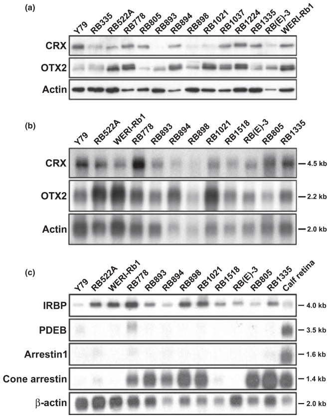
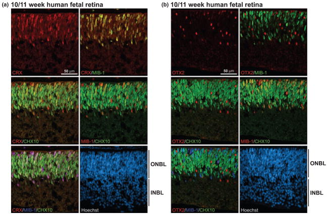
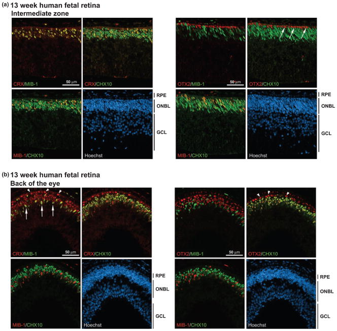
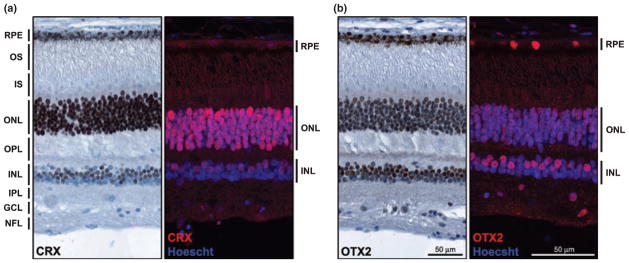
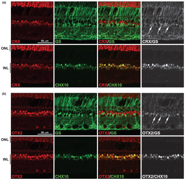
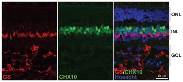
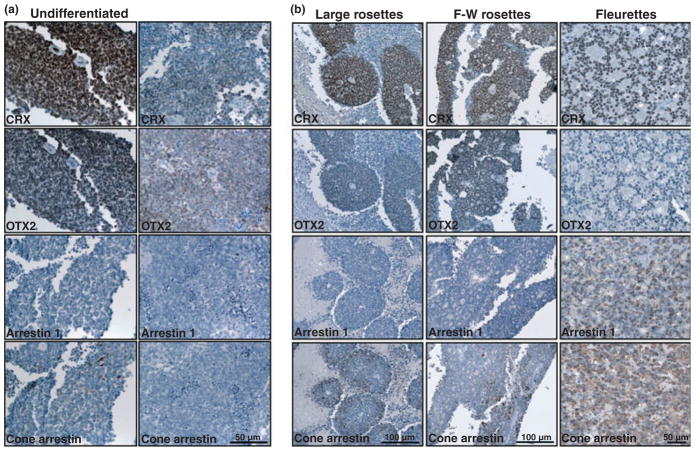
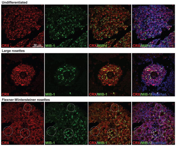
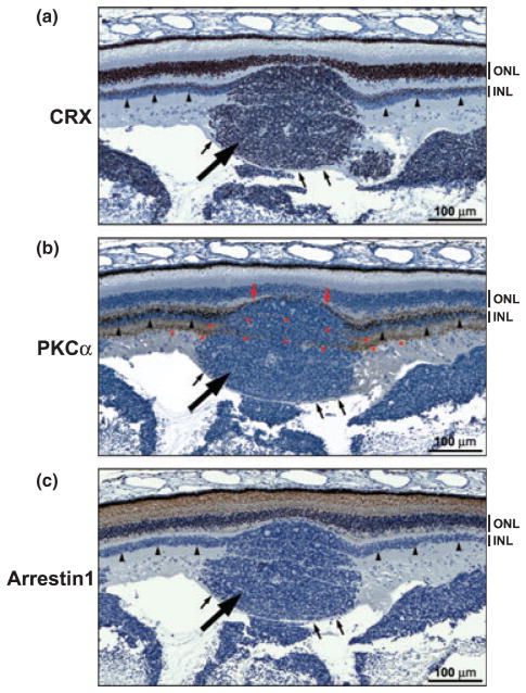
The histogenesis of retinoblastoma tumors remains controversial, with the cell-of-origin variably proposed to be an uncommitted retinal progenitor cell, a bipotent committed cell, or a cell committed to a specific lineage. Here, we examine the expression of two members of the orthodenticle family implicated in photoreceptor and bipolar cell differentiation, cone-rod homeobox, CRX, and orthodenticle homeobox 2, OTX2, in normal human retina, retinoblastoma cell lines and retinoblastoma tumors. We show that CRX and OTX2 have distinct expression profiles in the developing human retina, with CRX first expressed in proliferating cells and cells committed to the bipolar lineage, and OTX2 first appearing in the photoreceptor lineage. In the mature retina, CRX levels are highest in photoreceptor cells whereas OTX2 is preferentially found in bipolar cells and in the retinal pigmented epithelium. Both CRX and OTX2 are widely expressed in retinoblastoma cell lines and in retinoblastoma tumors, although CRX is more abundant than OTX2 in the differentiated elements of retinoblastoma tumors such as large rosettes, Flexner-Wintersteiner rosettes and fleurettes. Widespread expression of CRX and OTX2 in retinoblastoma tumors and cell lines suggests a close link between the cell-of-origin of retinoblastoma tumors and cells expressing CRX and OTX2.


Figures

Fig. 1
Fig. 1
(a) CRX and OTX2 expression in retinoblastoma cell lines. Western blot analysis of retinoblastoma cell lines using anti-CRX and anti-OTX2 antibodies. Fifty μg of whole cell lysates were loaded in each lane and electrophoresed in a 12% polyacrylamide-SDS gel. Proteins were transferred to PVDF membranes and sequentially immunostained with rabbit anti-CRX, followed by rabbit anti-OTX2 antibody (after stripping the blot), and goat anti-actin antibody. The signal was detected using the ECL or Immobilon detection reagents. (b) Northern blot analysis of CRX and OTX2 in retinoblastoma cell lines. Poly(A)+ RNA was isolated from 12 retinoblastoma cell lines. Two μg poly(A)+ RNA were loaded in each lane. The RNA was electrophoresed in an agarose-formaldehyde gel and the RNA transferred to a nitrocellulose filter. The filter was sequentially hybridized with 32P-labeled CRX, OTX2 and β-actin cDNAs. Sizes of the transcripts are indicated on the side. (c) Northern blot analysis of CRX/OTX2 target genes in retinoblastoma cell lines. Two μg poly(A)+ RNA from each of the indicated retinoblastoma cell lines and calf retina were electrophoresed in a 1.5% agarose-formaldehyde gel and transferred to a nitrocellulose filter. The filter was sequentially hybridized with 32P-labeled IRBP, PDEB, arrestin1, cone arrestin and β-actin cDNAs. The latter served as the loading control. Sizes of the transcripts are indicated on the side.
Fig. 2
Fig. 2
CRX and OTX2 expression in normal human fetal retina at 10– 11 weeks gestation. Tissue sections from paraformaldehyde-fixed human fetal retina at 10/11 weeks gestation were triple-stained with either rabbit anti-CRX antibody (a) or rabbit anti-OTX2 antibody (b), mouse MIB-1 antibody and sheep anti-CHX10 antibody. The signal was detected using donkey anti-rabbit Alexa 555 secondary antibody (CRX, OTX2), donkey anti-mouse Alexa 647 secondary antibody (MIB-1) and donkey anti-sheep Alexa 488 secondary antibody (CHX10). CRX/MIB-1, CRX/CHX10 and MIB-1/CHX10 co-stained cells range from orange to yellow depending on the relative intensity of the red and green signals. INBL, inner neuroblastic layer; ONBL, outer neuroblastic layer.
Fig. 3
Fig. 3
CRX and OTX2 expression in normal human fetal retina at 13 weeks gestation. Tissue sections from paraformaldehyde-fixed human fetal retina at 13 weeks gestation were triple-stained with either rabbit anti-CRX antibody or rabbit anti-OTX2 antibody, MIB-1 antibody and sheep anti-CHX10 antibody. Two different regions of the retina are represented: (a) intermediate differentiation zone and (b) back of the eye, the most differentiated part of the retina. The signal was detected using donkey anti-rabbit Alexa 555 secondary antibody (CRX, OTX2), donkey anti-mouse Alexa 647 secondary antibody (MIB-1) or donkey anti-sheep Alexa 488 secondary antibody (CHX10). The arrows in (a) point to the single layer of OTX2-positive photoreceptor cells. The arrows in (b) point to the CRX/MIB-1-positive cells and the arrowheads indicate the CRX-positive photoreceptor cells. Note that there is a layer of OTX2-expressing RPE cells immediately next to the OTX2-positive photoreceptor cells. RPE, retinal pigmented epithelium; ONBL, outer neuroblastic layer; GCL, ganglion cell layer.
Fig. 4
Fig. 4
CRX and OTX2 expression in mature retina. Tissue sections from a formalin-fixed paraffin-embedded eye from a 4-year-old child with retinoblastoma (patient No. 35917) were immunostained with anti- CRX antibody (a) or anti-OTX2 antibody (b), and analyzed by immunohistochemistry (brown signal) or immunofluorescence microscopy (red signal). The sections shown are from unaffected, apparently normal retina. For immunohistochemical analysis, the tissue section was counter-stained with the nuclear stain hematoxylin (blue) and the CRX or OTX2 signal detected using the DakoCytomationEnVision+ system. For immunofluorescence analysis, the tissue section was counter-stained with the nuclear stain Hoechst 33342 (blue) and CRX- and OTX2-positive cells (red) detected using Alexa 555-conjugated donkey anti-rabbit secondary antibody. NFL, nerve fiber layer; GCL, ganglion cell layer; IPL, inner plexiform layer; INL, inner nuclear layer; OPL, outer plexiform layer; ONL, outer nuclear layer (photoreceptor); IS, inner segments; OS, outer segments; RPE, retinal pigmented epithelium.
Fig. 5
Fig. 5
Co-immunostaining of CRX and OTX2 with markers of bipolar and Müller glial cells. A tissue section from an unaffected region of the eye of patient 35917 was triple-stained with rabbit anti-CRX antibody (a) or rabbit anti-OTX2 antibody (b), mouse anti-glutamine synthetase (GS) antibody and sheep anti-CHX10 antibody. Signals were detected with Alexa 555-conjugated donkey anti-rabbit (CRX, OTX2), Alexa 647-conjugated donkey anti-mouse (GS) and Alexa 488-conjugated donkey anti-sheep secondary antibodies (CHX10). Merging the CRX (a) or OTX2 (b) (red) and CHX10 (green) signals demonstrates their co-expression in bipolar cells (indicated by the yellow color). Merging the CRX (a) or OTX2 (b) (red) and GS (green) signals demonstrates their co-expression in Müller glial cells. Non-co-localized signals have been removed in the panels on the right in order to highlight sites of co-expression. Arrows point to CRX/GSand OTX2/GS-positive cells. INL, inner nuclear layer; ONL, outer nuclear layer (photoreceptor).
Fig. 6
Fig. 6
GS and CHX10 localization in normal human fetal retina at 7 months gestation. Slides containing tissue sections from normal human fetal retina at 7 months gestation were co-immunostained with anti- GS antibody (red) and CHX10 antibody (green). The merged figure demonstrates that there is little or no overlap in the staining pattern of GS and CHX10 in Müller glial cells. GCL, ganglion cell layer; INL, inner nuclear layer; ONL, outer nuclear layer.
Fig. 7
Fig. 7
Expression of CRX, OTX2, arrestin1 and cone arrestin in retinoblastoma tumors. (a) Tissue sections from two undifferentiated regions of a retinoblastoma tumor (patient 48292) were immunostained with anti-CRX antibody, anti-OTX2 antibody, anti-arrestin1 antibody and anti-cone arrestin antibody. Strong staining (brown color) is observed for both anti-CRX and OTX2 antibodies. No signal was detected using the anti-arrestin1 antibody whereas some staining was observed with anti-cone arrestin in the tumor on the left. (b) Tissue sections from differentiated regions of retinoblastoma tumors were immunostainined with anti-CRX antibody, anti-OTX2 antibody, anti-arrestin1 antibody and anti-cone arrestin antibody. Regions containing large rosettes (patient 15884) were strongly stained with anti-CRX antibody and moderately stained with anti-OTX2 and anti-cone arrestin antibody. Regions containing Flexner-Wintersteiner (F-W) rosettes (patient 45569) were strongly stained with anti-CRX antibody and moderately stained with anti-OTX2 and anti-cone arrestin antibody. Regions containing fleurettes (patient 48842) were strongly stained with anti-CRX antibody, anti-arrestin1 antibody and anti-cone arrestin antibody.
Fig. 8
Fig. 8
Co-staining of CRX and MIB-1 in retinoblastoma tumors. Tissue sections from retinoblastoma tumors (undifferentiated tumor from patient 48292 – top panels; large rosettes from patient 48292 – middle panels; Flexner-Wintersteiner rosettes from patient 45569 – bottom panels), were co-stained with anti-CRX and MIB-1 antibodies. Positive signals were detected using Alexa 488-conjugated donkey anti-rabbit (CRX) and Alexa 555-conjugated donkey anti-mouse (MIB-1) secondary antibodies. Sections were counter-stained with Hoescht 33342 to visualize nuclei and mounted with FluorSave reagent. The yellow color in CRX/MIB-1 merged panels demonstrates co-immunostaining. Although CRX-positive cells are more abundant than MIB-1-positive cells, the great majority of MIB-1-positive cells express CRX.
Fig. 9
Fig. 9
Expression of CRX, PKCα and arrestin1 in a transitory zone between retinal tissue and tumor. Tissue sections from patient 48842 were immunostained with anti-CRX15pep (a), anti-PKCα (b), and anti-arrestin1 (c) antibodies (DakoCytomationEnVision+ system) and counter-stained with hematoxylin. The tumor nodule is indicated by the thick arrow and the inner nuclear layer of the normal retinal tissue on either side of the tumor nodule is indicated by the arrowheads. The fiber layer overlaying the tumor is indicated by the small arrows. The PKCα-positive inner plexiform layer on either side of the tumor nodule (b) appears to extend through the tumor (as shown by the asterisks). INL, inner nuclear layer; ONL, outer nuclear layer.

References

    1. Ajioka I, Martins RA, Bayazitov IT, Donovan S, Johnson DA, Frase S, Cicero SA, Boyd K, Zakharenko SS, Dyer MA. Differentiated horizontal interneurons clonally expand to form metastatic retinoblastoma in mice. Cell. 2007;131:378–390. - PMC - PubMed
    1. Akagi T, Akita J, Haruta M, et al. Iris-derived cells from adult rodents and primates adopt photoreceptor-specific phenotypes. Invest Ophthalmol Vis Sci. 2005;46:3411–3419. - PubMed
    1. Bernardos RL, Barthel LK, Meyers JR, Raymond PA. Late-stage neuronal progenitors in the retina are radial Muller glia that function as retinal stem cells. J Neurosci. 2007;27:7028–7040. - PMC - PubMed
    1. Bibb LC, Holt JK, Tarttelin EE, Hodges MD, Gregory-Evans K, Rutherford A, Lucas RJ, Sowden JC, Gregory-Evans CY. Temporal and spatial expression patterns of the CRX transcription factor and its downstream targets. Critical differences during human and mouse eye development. Hum Mol Genet. 2001;10:1571–1579. - PubMed
    1. Boatright JH, Borst DE, Peoples JW, Bruno J, Edwards CL, Si JS, Nickerson JM. A major cis activator of the IRBP gene contains CRX-binding and Ret-1/PCE-I elements. Mol Vis. 1997;3:15. - PubMed

Publication types

MeSH terms

Grants and funding