. 2009 Aug 17:10:58.
doi: 10.1186/1471-2121-10-58.

Small G proteins in peroxisome biogenesis: the potential involvement of ADP-ribosylation factor 6

Affiliations

Small G proteins in peroxisome biogenesis: the potential involvement of ADP-ribosylation factor 6

Erin A Anthonio et al. BMC Cell Biol. .

Abstract

Background: Peroxisomes execute diverse and vital functions in virtually every eukaryote. New peroxisomes form by budding from pre-existing organelles or de novo by vesiculation of the ER. It has been suggested that ADP-ribosylation factors and COPI coatomer complexes are involved in these processes.

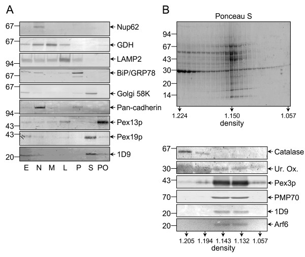
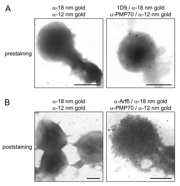
Results: Here we show that all viable Saccharomyces cerevisiae strains deficient in one of the small GTPases which have an important role in the regulation of vesicular transport contain functional peroxisomes, and that the number of these organelles in oleate-grown cells is significantly upregulated in the arf1 and arf3 null strains compared to the wild-type strain. In addition, we provide evidence that a portion of endogenous Arf6, the mammalian orthologue of yeast Arf3, is associated with the cytoplasmic face of rat liver peroxisomes. Despite this, ablation of Arf6 did neither influence the regulation of peroxisome abundance nor affect the localization of peroxisomal proteins in cultured fetal hepatocytes. However, co-overexpression of wild-type, GTP hydrolysis-defective or (dominant-negative) GTP binding-defective forms of Arf1 and Arf6 caused mislocalization of newly-synthesized peroxisomal proteins and resulted in an alteration of peroxisome morphology.

Conclusion: These observations suggest that Arf6 is a key player in mammalian peroxisome biogenesis. In addition, they also lend strong support to and extend the concept that specific Arf isoform pairs may act in tandem to regulate exclusive trafficking pathways.


Figures

Figure 1
Figure 1
Phenotypic analysis of various S. cerevisiae (deletion) strains. Serial dilutions of wild-type (WT) yeast cells (strain BY4741) and yeast cells deficient in Pex5p (Δpex5), Ypt8 (Δypt8), Arf1 (Δarf1), Arf2 (Δarf2), or Arf3 (Δarf3) expressing EGFP-PTS1 were spotted onto plates with oleate as a sole carbon source. (A) The plates were subsequently incubated at 30°C for seven days, and oleate consumption was scored by halo formation. (B) Two days later, the subcellular distribution pattern of EGFP-PTS1 was visualized by fluorescence microscopy. The scale bar represents 5 μm. (C) The number of peroxisomes per cell was counted in randomly selected cells. The mean number of peroxisomes per cell is indicated by an asterisk. At least 100 glucose-grown and 400 oleate-grown cells were scored.
Figure 2
Figure 2
Arf6 copurifies with the peroxisomal membrane fractions. (A) Rat liver homogenates were fractionated into a postnuclear (E), a nuclear (N), a heavy mitochondrial (M), a light mitochondrial (L), a microsomal (P), and a cytosolic (S) fraction. A peroxisome-enriched fraction (PO) was obtained by centrifugation of the L-fraction on a Nycodenz step gradient (see Methods). Equal amounts of protein were subjected to SDS-PAGE, transferred to nitrocellulose, and immunoblotted with antibodies raised against the nuclear pore complex protein p62 (Nup62), mitochondrial glutamate dehydrogenase (GDH), lysosomal-associated membrane protein 2 (LAMP2), the ER-resident chaperone immunoglobulin binding protein (BiP/GRP78), the microtubule-binding peripheral Golgi membrane 58 kDa protein (Golgi 58 K), the plasma membrane protein pan-cadherin (Pan-cadherin), the peroxisomal membrane protein Pex13p (Pex13p), peroxisomal biogenesis factor 19 (Pex19p), or a linear epitope found in Arf proteins (1D9). Note that the majority of Golgi 58 K is soluble after fractionation, and that Pex19p is a predominantly cytosolic, partly peroxisomal protein. (B) Six milligrams of total protein from the peroxisomal fraction was processed for floatation centrifugation in an alkaline sucrose gradient (see Methods). The fractions were collected from the bottom, processed for SDS-PAGE, transferred to a nitrocellulose membrane, and stained for total protein with Ponceau S (upper panel). A select set of fractions was immunoblotted with anti-Arf 1D9 or antibodies against Arf6 or peroxisomal matrix (catalase), core (urate oxidase), or membrane proteins (Pex3p, PMP70) (lower panels). The density (in g/ml) of the gradient fractions and the migration of the molecular mass markers (their masses expressed in kDa) are indicated.
Figure 3
Figure 3
Electron microscopic localization of 1D9-immunoreactive proteins in prestained epoxy-embedded preparations of rat liver peroxisomes. Nycodenz-purified rat liver peroxisomes were processed for pre-embedding double immunoelectron microscopy (see Methods). Rabbit anti-PMP70 (α-PMP70) and mouse monoclonal 1D9 antibodies were used as primary antibodies, and 12 nm gold-conjugated anti-rabbit IgG (α-12 nm gold) and 18 nm gold-conjugated anti-mouse IgG (α-18 nm gold) as labels. The primary antibodies were omitted from samples used as negative control (left column). The images were obtained from 75 nm (upper panels) and 300 nm thick (lower panels) sections. The original magnification was 27,800-fold; the scale bar represents 500 nm.
Figure 4
Figure 4
Electron microscopic localization of 1D9- and anti-Arf6-immunoreactive proteins in Nycodenz-purified peroxisomal fractions immobilized on poly-L-lysine-coated grids. (A) Prestained organellar fraction (see Methods). Rabbit anti-PMP70 (α-PMP70) and mouse monoclonal 1D9 were used as primary antibodies, and 12 nm gold-conjugated anti-rabbit IgG (α-12 nm gold) and 18 nm gold-conjugated anti-mouse IgG (α-18 nm gold) as labels. (B) Poststained organellar fraction (see Methods). Rabbit anti-PMP70 (α-PMP70) and mouse anti-Arf6 (α-Arf6) were used as primary antibodies, and the same secondary antibodies were used as in panel A. Negative controls were included in parallel in which the primary antibodies were omitted (left panels). The original magnification was 60,000-fold (upper panels), 27,800-fold (lower left panel), or 46,460 fold (lower right panel); the scale bar represents 500 nm.
Figure 5
Figure 5
Organellar distribution of 1D9- and anti-PMP70-immunoreactive proteins in enriched peroxisomal rat liver fractions. Percoll-, Nycodenz- and Percoll/Nycodenz-purified peroxisomal fractions were processed for pre-embedding double immunoelectron microscopy using anti-Arf 1D9 (18 nm gold grains) and anti-PMP70 (12 nm gold grains) antibodies (see legend to Figure 3). Epon-embedded fractions were quantitatively analyzed for the density of labeling on organellar membranes. The results are expressed as relative specific labeling (R.S.L.) versus percentage of total cell organelles. R.S.L. is hereby defined as the percentage of total gold particles present on a particular organelle divided by the corresponding percentage of total organelles. R.S.L. values greater than one indicate enrichment of the labeling in that specific fraction. The combined total number of cell organelles (x-axis) and gold particles (y-axis) is indicated. Note that the purity of the peroxisomal Percoll, Nycodenz, and Percoll/Nycodenz fractions was 60%, 70%, and 77%, respectively. Abbreviations: PO, peroxisomes (vesicular structures labeled with anti-PMP70 antibodies); MT, mitochondria (double membrane-bound organelles with cristae); O, other vesicles (vesicular structures which could not be unambiguously identified as peroxisomes or mitochondria).
Figure 6
Figure 6
Arf6 is not essential for peroxisome biogenesis in fetal mouse hepatocytes from control and clofibrate-treated pregnant mice. (A) Protein (25 μg), present in liver postnuclear supernatants from control (-CF) and clofibrate-treated (+CF) pregnant Arf6+/- mice (see Methods), was subjected to SDS-PAGE, transferred to PVDF, and immunoblotted with antibodies against cytochrome P450 4A (Cyt P450; a non-peroxisomal clofibrate-inducible enzyme), the L-specific peroxisomal multifunctional protein (MFP1; a clofibrate-inducible enzyme), peroxisomal thiolase (thiol; a clofibrate-inducible enzyme), or catalase (cat; a peroxisomal enzyme not induced by clofibrate). The asterisk indicates the migration of a nonspecific, immunoreactive protein. (B, C) Primary hepatocytes from mouse embryos (13.5 days) of Arf6+/+ and Arf6-/- littermates from control and clofibrate-treated pregnant Arf6+/- mice were isolated, seeded on collagen-coated cover glasses, cultured for 12 hours, and processed for indirect immunofluorescence microscopy. (B) Representative pictures showing that ARF6 ablation does not alter the localization of Pex14p, a peroxisomal membrane protein. The scale bar represents 10 μm. (C) The number of peroxisomes (per cell section; n > 15) is not substantially altered in wild-type and Arf6-/- cells. The error bars indicate the standard deviation.
Figure 7
Figure 7
Effect of co-overexpression of Arf1 and Arf6 variants on peroxisomal and mitochondrial protein import in PtK2 cells. (A) Ptk2 cells were transiently transfected with plasmids coding for no protein (/), Arf1WT-HA (1), Arf1T31N-HA (1-), or Arf1Q71L-HA (1+) and/or a bicistronic plasmid encoding EGFP-PTS1 together with no other protein (-), non-tagged Arf6WT (6), Arf6T27N (6-), or Arf6Q67L (6+). After 36 hours, the cells were fixed and processed for fluorescence analysis. The subcellular localization of EGFP-PTS1 was determined by its punctate (peroxisomal) or diffuse staining pattern in at least 250 cells, and the results were quantified. (B) Representative images of the subcellular distribution pattern of EGFP-PTS1, HsPMP34-Myc-His, and HsLK2-Myc-His in Ptk2 cells co-overexpressing Arf1T31N and Arf6T27N. Note that the latter protein is encoded by the bicistronic expression vector coding for EGFP-PTS1, and a mislocalization of the reporter proteins is only observed in double-transfected cells. As (i) in mammalian cells the fluorescence intensity of EGFP-PTS1 is significantly higher upon mislocalization to the cytosol [77], and (ii) this increase may mask the (partial) association of this reporter protein with peroxisomes, insets are included in which the outlined regions are shown with moderately less intense green fluorescence signals. Scale bar: 20 μm.

Similar articles

Cited by

References

    1. Wanders RJ, Waterham HR. Biochemistry of mammalian peroxisomes revisited. Annu Rev Biochem. 2006;75:295–332. doi: 10.1146/annurev.biochem.74.082803.133329. - DOI - PubMed
    1. Mano S, Nishimura M. Plant peroxisomes. Vitam Horm. 2005;72:111–154. full_text. - PubMed
    1. Schrader M, Fahimi HD. Peroxisomes and oxidative stress. Biochim Biophys Acta. 2006;1763:1755–1766. doi: 10.1016/j.bbamcr.2006.09.006. - DOI - PubMed
    1. Klei I van der, Veenhuis M. Yeast and filamentous fungi as model organisms in microbody research. Biochim Biophys Acta. 2006;1763:1364–1373. doi: 10.1016/j.bbamcr.2006.09.014. - DOI - PubMed
    1. Geuze HJ, Murk JL, Stroobants AK, Griffith JM, Kleijmeer MJ, Koster AJ, Verkleij AJ, Distel B, Tabak HF. Involvement of the endoplasmic reticulum in peroxisome formation. Mol Biol Cell. 2003;14:2900–2907. doi: 10.1091/mbc.E02-11-0734. - DOI - PMC - PubMed

Publication types

MeSH terms