. 2009 Aug;17(2):175-86.
doi: 10.1016/j.devcel.2009.06.017.

Integrin-alpha9 is required for fibronectin matrix assembly during lymphatic valve morphogenesis

Affiliations

Integrin-alpha9 is required for fibronectin matrix assembly during lymphatic valve morphogenesis

Eleni Bazigou et al. Dev Cell. 2009 Aug.

Abstract

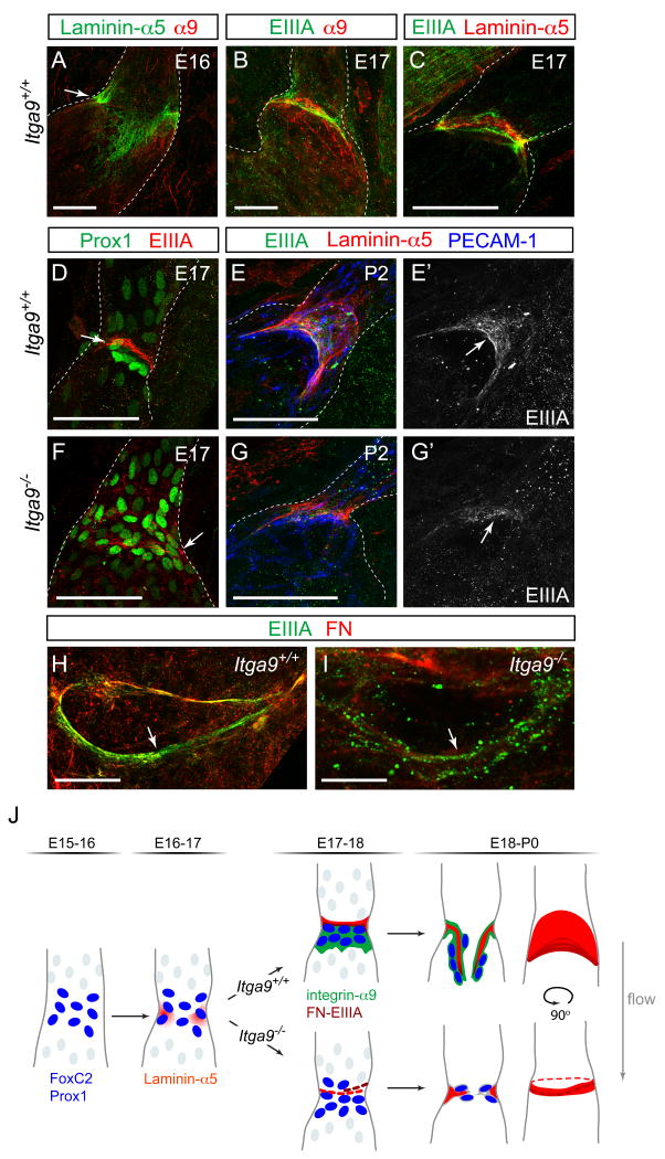
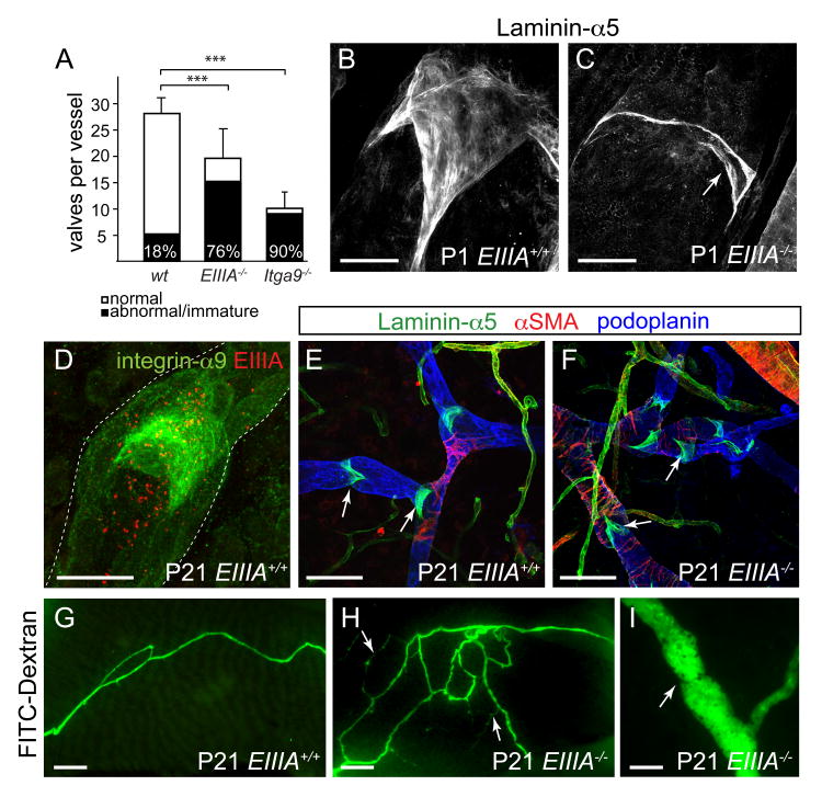
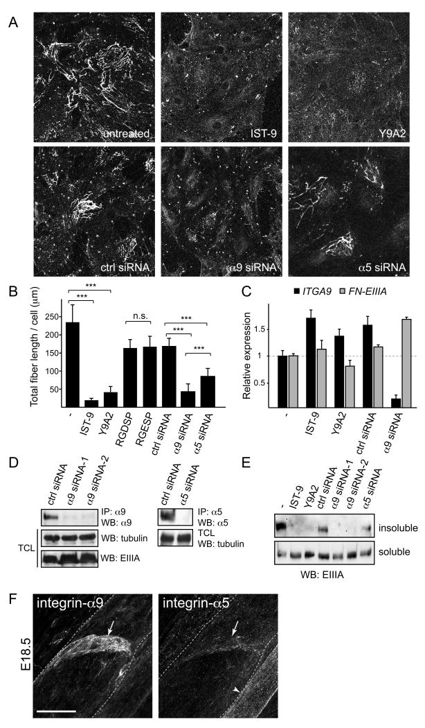
Dysfunction of lymphatic valves underlies human lymphedema, yet the process of valve morphogenesis is poorly understood. Here, we show that during embryogenesis, lymphatic valve leaflet formation is initiated by upregulation of integrin-alpha9 expression and deposition of its ligand fibronectin-EIIIA (FN-EIIIA) in the extracellular matrix. Endothelial cell-specific deletion of Itga9 (encoding integrin-alpha9) in mouse embryos results in the development of rudimentary valve leaflets characterized by disorganized FN matrix, short cusps, and retrograde lymphatic flow. Similar morphological and functional defects are observed in mice lacking the EIIIA domain of FN. Mechanistically, we demonstrate that in primary human lymphatic endothelial cells, the integrin-alpha9-EIIIA interaction directly regulates FN fibril assembly, which is essential for the formation of the extracellular matrix core of valve leaflets. Our findings reveal an important role for integrin-alpha9 signaling during lymphatic valve morphogenesis and implicate it as a candidate gene for primary lymphedema caused by valve defects.


Figures

Figure 1
Figure 1. Expression of integrin-α9 in mature and developing lymphatic valves
(A, B) Immunofluorescence staining of adult ear skin with antibodies against integrin-α9 (green), FoxC2 (red) and α-smooth muscle actin (α-SMA, blue). Arrow in (B) points to a luminal valve. (C-E) Development of mesenteric lymphatic vessels. Whole-mount X-Gal staining of mesenteric lymphatic vessels from Vegfr3lz/+ embryos. The tissues were taken from embryos at the indicated ages (E16.5-E18.5). (F-K) Immunofluorescence staining of developing mesenteric lymphatic vessels of E16.5 (F, G), E17.5 (H, I) and E18.5 (J, K) with antibodies against integrin-α9 (green), Prox1 (F, red) or FoxC2 (H, J, red) and LYVE-1 (F, H, J, blue). Arrowhead in (F, G) points to a blood vessel, the smooth muscle coverage of which is positive for integrin-α9 staining. Arrows point to clusters of cells expressing high levels of Prox1 and FoxC2. Scale bars; A, B, H-K: 50 μm, C-G: 1 mm.
Figure 2
Figure 2. Abnormal valves in Itga9 deficient mice
(A-B′) Luminal valves in chyle-filled mesenteric lymphatic vessels of wild-type (A, A′) and Itga9 mutant mice (B, B′). Note the difference in the shape of a wild-type in comparison to a mutant valve (A′, B′, arrows in A, B) and leakage of chyle from the mutant vessels (arrowhead in B). BF = bright field. (C, D) PECAM-1 immunohistochemistry of P5 mesenteric vessels and luminal valves (arrow) in wild-type (C) and Itga9-/- (D) mice. (E) Quantification of the number of luminal valves in P5 wild-type and Itga9-/- mesenteric lymphatic vessels (mean ± s.d., n = 4 animals per genotype, 3 vessels each). Black bar = normal V-shaped valves; white bar = abnormal valves with ring appearance. *** p < 0.0001 (Mann-Whitney test). (F) Schematic representation of luminal valves (arrows) in the collecting lymphatic vessels of Itga9+/+ and Itga9-/- mice. (G-J) Transmission electron micrographs of wild-type (G, I) and Itga9-/- (H, J) valves in mesenteric lymphatic vessels of P6 mice. Arrows in (G, H) point to the matrix core (red) anchored into the vessels wall, arrowheads mark the free edges of the valve leaflets. (I) shows the valve leaflet with a connective tissue core (red). Note the rudimentary (arrows in H) or absent (J) matrix core in the mutant valves and the gaps in between the two endothelial sheets (red asterisks in J). (K) Schematic representation of luminal valves in the Itga9+/+ and Itga9-/- mice. Matrix core is indicated in red. Scale bars; A, B: 100 μm, C, D: 50 μm, G, H: 10 μm, I, J: 1 μm.
Figure 3
Figure 3. Defective lymphatic drainage in Itga9 deficient mice
(A, B) Visualisation of dermal collecting vessels following injection of FITC-dextran into the footpads of P6 wild-type (A) and Itga9-/- mice (B). Note the presence of an abnormal vessel network (arrowheads in B) and a valve in a vessel branch point in the Itga9 mutants (arrow in B). (C, D) FITC-lectin (LEL) staining of the valves in dermal lymphatic vessels following footpad injection. No valve leaflets are seen in the Itga9 mutant (D). Arrows in (C) point to the two valve leaflets seen from the 90° angle when compared to Figure 2A, D. (E) Schematic representation of a side view of luminal valves as visualized by FITC-LEL staining in the collecting lymphatic vessels of Itga9+/+ (left) and Itga9-/- (right) mice. Arrows indicate the direction of the flow. Scale bars; A, B: 100 μm, C, D: 50 μm.
Figure 4
Figure 4. Endothelial cell specific deletion of Itga9 during development and in mature valves
Lymphatic vessels of Tie2-Cre;Itga9lx/lx mouse (A-D), and of 4-OHT treated Itga9lx/+ (E-H) and VEcad-CreERT2; Itga9lx/lx (I-L) mice stained with antibodies against Laminin-α5 (red), integrin-α9 (green) and PECAM-1 (blue). Expression of integrin-α9 is detected in the vascular SMC (arrowhead in A, C, E, G, I, K) but is lost from the endothelial cells in Tie2-Cre;Itga9lx/lx (C) and from most endothelial cells of the valves of VEcad-CreERT2; Itga9lx/lx mutant animals (I, K, open arrowhead points to a single integrin-α9 expressing cell). Note the abnormal valve in Tie2-Cre;Itga9lx/lx mouse (arrow in B), which has undergone embryonic deletion of Itga9 allele, but intact valve leaflets (arrow in J) and the attachment of LECs on the leaflets in the VEcad-CreERT2; Itga9lx/lx mutant (arrow in L), which has undergone postnatal deletion of Itga9 allele, as compared to a control (arrows in F, G). Scale bars; A-L: 20 μm.
Figure 5
Figure 5. Development of lymphatic valve leaflets in wild-type and Itga9-/- mice
(A-C) Immunofluorescence staining of developing mesenteric lymphatic vessels of E16 (A) and E17 (B, C) wild-type embryo using antibodies against Laminin-α5, integrin-α9 and FN-EIIIA (colors as indicated). The dotted lines outline the vessels. (D-G) Immunolabeling of lymphatic valves in wild-type (D-E′) and Itga9-/- (F-G′) mesenteric vessels for Prox1 (green) and FN-EIIIA (red; E, F; at E17) or for Laminin-α5 (red), FN-EIIIA (green) and the endothelial marker PECAM-1 (blue; E, G; at P2). The arrows in (D, E′, F, G′) point to FN-EIIIA fibers. (H, I) View through the opening of the valve in P0 wild-type (H) and Itga9-/- (I) vessels, labelled for FN (red) and FN-EIIIA (green). Note the punctuate localization of FN-EIIIA in the Itga9-/- valve (arrow in I) compared to the fibrous staining in the wild-type (arrow in H). (J) Schematic model of lymphatic valve formation. Upregulation of Prox1 and FoxC2 transcription factors (blue nuclei) in lymphatic vessels define the positions of future valves. Deposition of extracellular matrix (red) containing Laminin-α5 and FN-EIIIA and re-orientation of cells expressing high levels of Prox1 and FoxC2 perpendicular to the vessel wall is followed by upregulation of integrin-α9 (green) on the outflow side of the future valve. Itga9-/- mice (below) display defective organization of the extracellular matrix and failure of leaflet formation. Scale bars; A-G: 50 μm, H, I: 10 μm.
Figure 6
Figure 6. Abnormal lymphatic valves in mice lacking the integrin-α9 ligand, FN-EIIIA
(A) Luminal valve numbers in newborn wild-type, Fn-EIIIA-/- and Itga9-/- mesenteric lymphatic vessels (mean ± s.d., n ≥ 4 animals per genotype, ≥ 2 vessels each, see Suppl. Table 1). The percentage of abnormal valves is indicated: *** p < 0.0001 (χ2 test). (B, C) Visualization of lymphatic valves in P1 wild-type (B) and Fn-EIIIA-/- (C) mesenteric vessels using antibodies against Laminin-α5. Note the incomplete development of the valve as evident by lack of leaflets (arrow in C) in the Fn-EIIIA-/- vessels. (D) Immunofluorescence staining of three weeks old ear skin for integrin-α9 (green) and EIIIA (red). (E, F) Dermal lymphatic vessels in the ears of three weeks old wild-type (E) and Fn-EIIIA-/- (F) mice labeled for Laminin-α5 (green), podoplanin (blue) and α-SMA (red). (G-I) FITC-dextran assay in three weeks old wild-type (G) and Fn-EIIIA-/- mice (H, I). Note the reflux of dye (arrows in H) and an abnormal valve (arrow in I) in the mutant skin. Scale bars; B, C: 20 μm, D:50μm, E, F, I: 100 μm, G, H: 400 μm.
Figure 7
Figure 7. Integrin-α9-EIIIA interaction regulates FN fibril assembly in primary human lymphatic endothelial cells
(A) FN fibrils in primary human lymphatic endothelial cells (LECs). Integrin-α9-EIIIA interaction was blocked using antibodies against EIIIA (IST-9) or integrin-α9β1 (Y9A2), or siRNA against integrin-α9 or -α5, and stained with EIIIA antibodies. (B) Quantification of FN fibrillogenesis in the LECs, in which integrin-α9-EIIIA interaction (IST-9, Y9A2, α9 siRNA) or integrin-α5/RGD-dependent integrin interactions (RGDSP peptide, α5 siRNA) were inhibited, in comparison to the control cells (untreated, ctrl siRNA or RGESP peptide). Data represent mean FN-EIIIA fiber length per cell (± s.d) from five randomly chosen view fields in two independent experiments. *** p< 0.003, n.s.= non-significant, p = 0.881 (Student T-test). (C) qPCR of ITGA9 and FN-EIIIA in human LECs. Data represent mean ± s.d. of triplicates. (D) siRNA mediated knock-down of integrin expression in primary human LECs. Western blot analysis of immunoprecipitated (IP) cell lysates using integrin-α9 or -α5 antibodies (upper panels). For the loading control, the total cell lysates (TCL) were blotted against α-tubulin and EIIIA. (E) Conversion of DOC-soluble FN fibrils into insoluble stable matrix. DOC-insoluble (upper panel) and -soluble matrix (lower panel) isolated from the LECs were separated in non-reducing SDS-PAGE and probed for EIIIA. (F) Immunofluorescent staining of wild type E18 mesenteric vessels using antibodies against integrin-α9 (left panel) and integrin-α5 (right panel). Note low levels of integrin-α5 expression in the valve (arrows) in comparison to strong staining in the blood vessel endothelia (arrowhead). Scale bar = 20 μm.

References

    1. Alitalo K, Tammela T, Petrova TV. Lymphangiogenesis in development and human disease. Nature. 2005;438:946–953. - PubMed
    1. Armstrong EJ, Bischoff J. Heart valve development: endothelial cell signaling and differentiation. Circ Res. 2004;95:459–470. - PMC - PubMed
    1. Astrof S, Crowley D, George EL, Fukuda T, Sekiguchi K, Hanahan D, Hynes RO. Direct test of potential roles of EIIIA and EIIIB alternatively spliced segments of fibronectin in physiological and tumor angiogenesis. Mol Cell Biol. 2004;24:8662–8670. - PMC - PubMed
    1. Avraamides CJ, Garmy-Susini B, Varner JA. Integrins in angiogenesis and lymphangiogenesis. Nat Rev Cancer. 2008;8:604–617. - PMC - PubMed
    1. Dumont DJ, Jussila L, Taipale J, Lymboussaki A, Mustonen T, Pajusola K, Breitman M, Alitalo K. Cardiovascular failure in mouse embryos deficient in VEGF receptor-3. Science. 1998;282:946–949. - PubMed

Publication types

MeSH terms