. 2009 Aug;17(2):199-209.
doi: 10.1016/j.devcel.2009.07.013.

Ret-dependent cell rearrangements in the Wolffian duct epithelium initiate ureteric bud morphogenesis

Affiliations

Ret-dependent cell rearrangements in the Wolffian duct epithelium initiate ureteric bud morphogenesis

Xuan Chi et al. Dev Cell. 2009 Aug.

Abstract

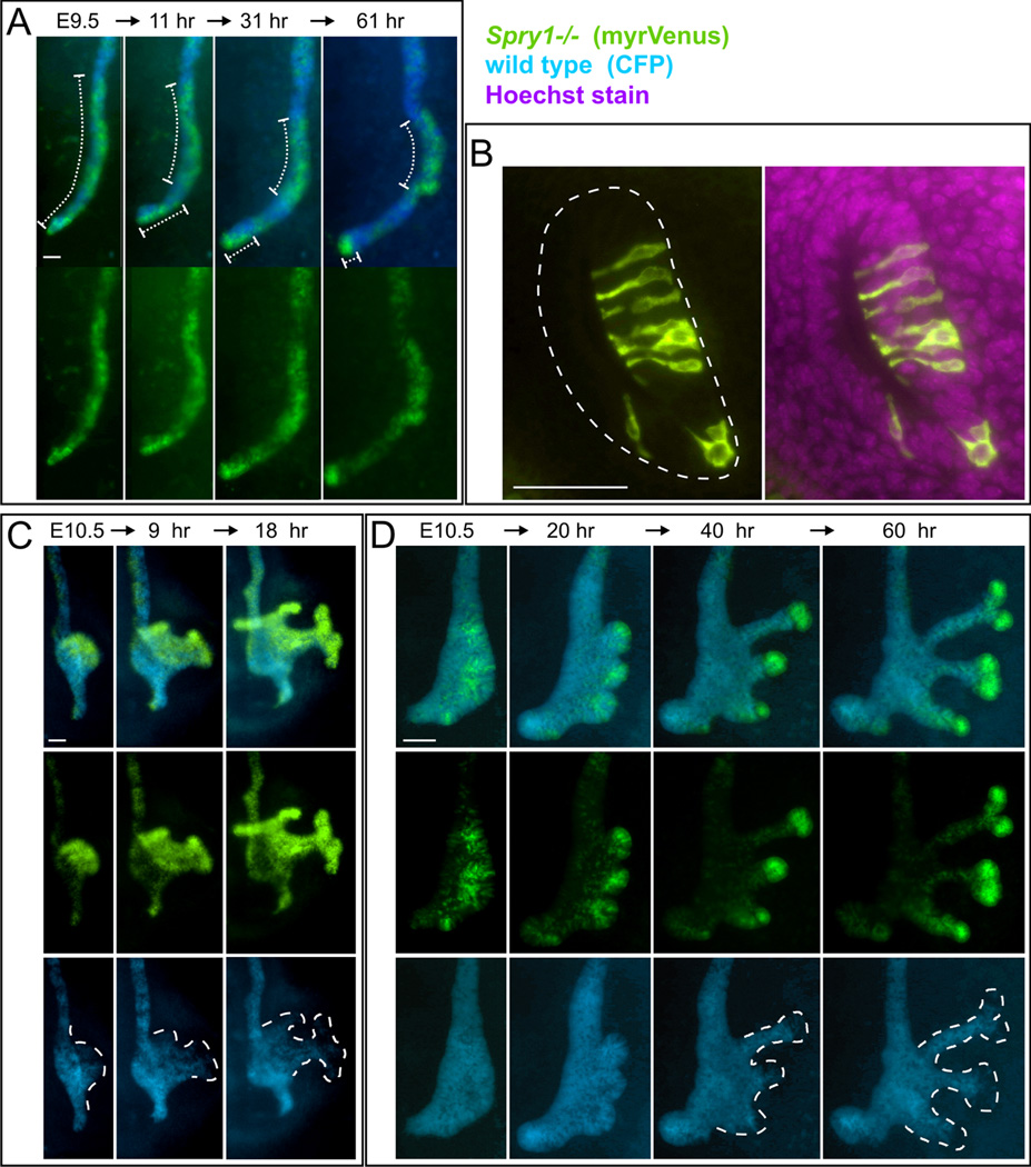
While the genetic control of renal branching morphogenesis has been extensively described, the cellular basis of this process remains obscure. GDNF/RET signaling is required for ureter and kidney development, and cells lacking Ret are excluded from the tips of the branching ureteric bud in chimeric kidneys. Here, we find that this exclusion results from earlier Ret-dependent cell rearrangements in the caudal Wolffian duct, which generate a specialized epithelial domain that later emerges as the tip of the primary ureteric bud. By juxtaposing cells with elevated or reduced RET activity, we find that Wolffian duct cells compete, based on RET signaling levels, to contribute to this domain. At the same time, the caudal Wolffian duct transiently converts from a simple to a pseudostratified epithelium, a process that does not require Ret. Thus, both Ret-dependent cell movements and Ret-independent changes in the Wolffian duct epithelium contribute to ureteric bud formation.


Figures

Figure 1
Figure 1. Behavior of wild type and Ret−/− cells during the formation, outgrowth and initial branching of the UB
(A) Ret−/− ES cells carrying Hoxb7/GFP were injected into WT blastocysts, which were generated by crossing Hoxb7/Cre (Yu et al., 2002) and R26R-CFP mice (Srinivas et al., 2001). Therefore, WD/UB cells derived from the Ret−/− ES cells expressed GFP, while those derived from the WT host expressed CFP. Dissected urogenital regions were examined in whole mount. (B) A chimeric WD at ~E9.5, before the beginning of UB formation. GFP+ and CFP+ cells appear randomly distributed. (C) at ~E10.0, the dorsal side of the caudal WD, where the UB will later emerge, becomes highly enriched for wild type (CFP+) cells (bracket). (D–E) As the UB emerges (~E10.5), it is composed almost entirely of WT cells. (F) As the UB elongates (~E11.0), WT cells generate the tip, while mutant cells follow behind and contribute to the trunk. (G–H) During the first UB branching event (~E11.5) mutant cells contribute to the proximal sides of the branches, while only WT cells form the tips. (I) Control chimeric UB generated using wild type (Ret+/+) ES cells, which contribute uniformly. Scale bars, 100 µM.
Figure 2
Figure 2. Spatial distribution and properties of mutant and wild type cells in the pre-budding Ret−/− ↔ WT chimeric Wolffian duct
(A–C) Whole mount images of a chimeric E10.5 Wolffian duct, in which the primary UB tip domain (right side, dorsal) is composed mainly of wild type (CFP+) cells, while the ventral WD (left side) is depleted of wild type cells. Asterisk marks the CND, also highly enriched in WT cells. Dotted lines in A show approximate planes of the sections in (D–E) and (F–G) (which show a different chimeric embryo). (D–G) show one section through the rostral WD (D–E) and one through the caudal WD (F–G) of an E10.5 chimera. The host embryo did not carry CFP, and the blue nuclear stain is Hoechst. The rostral WD has a random distribution of mutant (GFP+) and WT cells, while in the caudal WD, mutant cells are absent in the primary UB tip domain (dorsal region, on right) but enriched in the ventral region. Red stain in (D, F) is anti-pH3. (H) % of mutant or WT cells that were pH3+, in the rostral, caudal or entire WD of chimeric embryos (mean +/− SD). Asterisks indicates a significant difference (p<0.01) between % wild type cells that were pH3+ cells in rostral vs. caudal WD. However, the % of mutant vs. WT pH3+ cells was not significantly different. (I – J) E-cadherin staining (red) of a chimeric WD shows similar expression in WT and mutant (GFP+) cells. GFP was detected by fluorescence (A–C) or anti-GFP (other panels). Scale bars, 20 µM.
Figure 3
Figure 3. The caudal Wolffian duct forms a transiently pseudostratified epithelium, in both wild type and Ret −/− embryos
The WD and UB epithelium was visualized using the Hoxb7/myr-Venus transgene. (A–E’) Wild type and Ret−/− Wolffian ducts at E10.0. (A) Whole mount image of WT Wolffian duct. Yellow lines indicate approximate planes of the sections at right, which show rostral WD from a wild type (B) or Ret−/− (D) embryo, and pseudostratified caudal WD from a wild type (C, C’) or Ret−/− (E, E’) embryo. B–E were stained with anti-GFP to detect myr-Venus (green) and anti-pH3 (red). (C’) and (E’) Hoechst nuclear stain of the sections in C and E. (F) Wild type UB at E11.5, in whole mount. Yellow line indicates approximate plane of section in (G). (H) Wild type UB at ~E14.5, in whole mount. Yellow box shows approximate area of the optical section through a terminal UB branch in (I). (J) Schematic diagram of the formation of pseudostratified epithelium preceding ureteric budding, and reversion to a simple epithelium by E14.5. Scale bars 100 µM in A, F, H, 20 µM in other panels.
Figure 4
Figure 4. Wild type cells in the chimeric Ret−/− ↔ WT Wolffian duct converge to form the primary UB tip domain
(A–B) Culture of a Ret−/−↔WT chimera (~E10.0) in which formation of the primary UB tip domain was visualized (Movie 1). (A) shows CFP and GFP images merged, revealing interspersion of WT (CFP+) and mutant (GFP+) cells in the Wolffian duct at 0 hr, but enrichment of WT cells at the primary UB tip domain (arrow) and common nephric duct (CND, *) by 24 hr. (B) shows the CFP channel, revealing rearrangement of WT cells to form the UB tip domain (brackets) and CND. (C–F) Culture of a Ret−/−↔WT chimera from E10.5 (Movies 2, 2a and 2b). The boxes in (C) indicate the areas enlarged in (D–F). At 0 hr (C, left), the normal primary UB tip domains had already formed (arrows). By 48 hr (C, right), the two normal UBs had grown out (arrows), and four ectopic buds (#1–4) had also emerged. (D) Enlargements from Movie 2 showing outgrowth, elongation and ampulla formation of a normal UB. Between 30 and 48 hr, CFP+ cells from the UB tip give rise to part of the trunk, consistent with lineage analyses (Shakya et al., 2005). (E–F) Enlargements from Movies 2a and 2b showing cell movements preceding evagination of buds #1–4. The CFP+ wild type cells, which are initially dispersed (brackets), converge into clusters before evaginating as the UB tips. Scale bars,100 µM.
Figure 5
Figure 5. In Spry1−/−↔WT chimeras, Spry1−/− cells preferentially populate the UB tip domain while wild type cells are excluded
(A) Movements of Spry1−/− cells in the Wolffian duct during culture of a Spry1−/−↔WT chimera isolated at ~E9.5 (produced by injecting Spry1−/−, Hoxb7/myr-Venus ES cells into Hoxb7/Cre;R26R-CFP blastocysts – see Figure 1A). The chimeric Wolffian duct is shown at four time points. Brackets indicate the changing distribution of Spry1−/− cells as they converge into two clusters, one at the site of future ureteric budding, and one at the CND. (B) In a section through the caudal Wolffian duct of a Spry1−/−↔WT chimera at ~E10.0, Spry1−/− cells preferentially populate the primary UB tip domain on the dorsal (right) side. (C–D) show the Wolffian ducts and ureteric buds of two Spry1−/−↔WT chimeras cultured from ~E10.5. In both cases, Spry1−/− cells predominate over WT cells to form the UB tips (dashed lines). See Movie 3 and Movie 4. Scale bars 100 µM in A, C, D, 50 µM in B.
Figure 6
Figure 6. In Ret-hypomorphic host embryos, unlike normal hosts, Ret−/− cells frequently contribute to the UB tips
Ret−/− ES cells (A–H) or control Ret+/− ES cells (I–J), both carrying Hoxb7/GFP, were injected into normal (left column) or Ret-hypomorphic blastocysts (right column). Kidneys were excised at E11.5 (A–D) or E12.5 (E–J) and cultured for 24 hr. The kidneys were stained with anti-cytokeratin (red) to visualize the UB epithelium (as host embryos did not carry the CFP gene), and the ES cell contribution was detected with anti-GFP (green). Brackets in C, D, G and H indicate Ret−/− cells in the UB tips. Insets in G and H show magnified images of the UB tips. Note that the presence of many Ret+/− cells in (J) corrected the branching defect seen in Ret-hypomorphic kidneys (G–H). Scale bars 100 µM.
Figure 7
Figure 7. Independent cell movements and heterogeneous Ret signaling in the wild type Wolffian duct
(A–C) diagram illustrating rearrangement of WT (blue) and mutant (green) cells in Ret −/−↔WT chimeras. Gray ovals represent metanephric mesenchyme. The initially dispersed WT cells (A) converge along the long axis of the Wolffian duct and also move dorsally (yellow arrows), to form the primary UB tip domain (B). When the UB grows out, WT cells lead, forming the tip, while Ret−/− cells follow (C, red arrows). (D–J) transverse sections of ~E10.0–10.5 embryos. (D) Ret mRNA is expressed uniformly in cells of the Wolffian duct (arrows). Dotted circles indicate GDNF-expressing MM. (E) In WT (37 somite) embryo, anti-dp-Erk specifically stains the WD domain that will form the UB tip (arrows; compare to Figure 2F,G). (F) In a Ret−/− embryo, the WD is dp-Erk-negative. (G–J) Wolffian duct sections (34–36 somite WT embryos) showing heterogeneous dp-Erk staining. (I) and (J) are caudal and (G) and (H) more rostral sections. (H) and (J) were counterstained with Hematoxylin. (K–L) Independent movements of neighboring cells during budding in non-chimeric Wolffian ducts. A random subset of duct cells was YFP-labeled by Tamoxifen treatment at E7.5 (K) or E9.5 (L) of RetCreERT2/+; R26RYFP/+ embryos (in which all cells have the same Ret+/− genotype). Urogenital regions were cultured from ~E10.5 for indicated number of hrs. In each sequence, several YFP+ cells enter the tip of a forming UB (yellow brackets) while other neighboring YFP+ cells remain behind in the WD (red brackets).Scale bars, 25 µM.

References

    1. Affolter M, Bellusci S, Itoh N, Shilo B, Thiery JP, Werb Z. Tube or not tube: remodeling epithelial tissues by branching morphogenesis. Dev Cell. 2003;4:11–18. - PubMed
    1. Affolter M, Caussinus E. Tracheal branching morphogenesis in Drosophila: new insights into cell behaviour and organ architecture. Development. 2008;135:2055–2064. - PubMed
    1. Basson MA, Akbulut S, Watson-Johnson J, Simon R, Carroll TJ, Shakya R, Gross I, Martin GR, Lufkin T, McMahon AP, et al. Sprouty1 is a critical regulator of GDNF/RET-mediated kidney induction. Dev Cell. 2005;8:229–239. - PubMed
    1. Basson MA, Watson-Johnson J, Shakya R, Akbulut S, Hyink D, Costantini FD, Wilson PD, Mason IJ, Licht JD. Branching morphogenesis of the ureteric epithelium during kidney development is coordinated by the opposing functions of GDNF and Sprouty1. Dev Biol. 2006;299:466–477. - PubMed
    1. Batourina E, Tsai S, Lambert S, Sprenkle P, Viana R, Dutta S, Hensle T, Wang F, Niederreither K, McMahon AP, et al. Apoptosis induced by vitamin A signaling is crucial for connecting the ureters to the bladder. Nat Genet. 2005;37:1082–1089. - PubMed

Publication types

MeSH terms