. 2009 Nov 15;47(10):1414-21.
doi: 10.1016/j.freeradbiomed.2009.08.008. Epub 2009 Aug 14.

Trypanosoma cruzi infection disturbs mitochondrial membrane potential and ROS production rate in cardiomyocytes

Affiliations

Trypanosoma cruzi infection disturbs mitochondrial membrane potential and ROS production rate in cardiomyocytes

Shivali Gupta et al. Free Radic Biol Med. .

Abstract

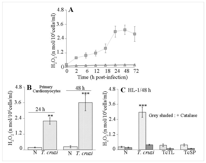
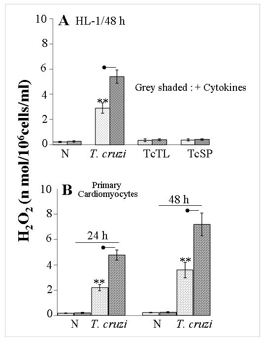
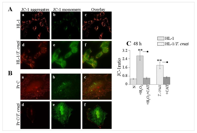
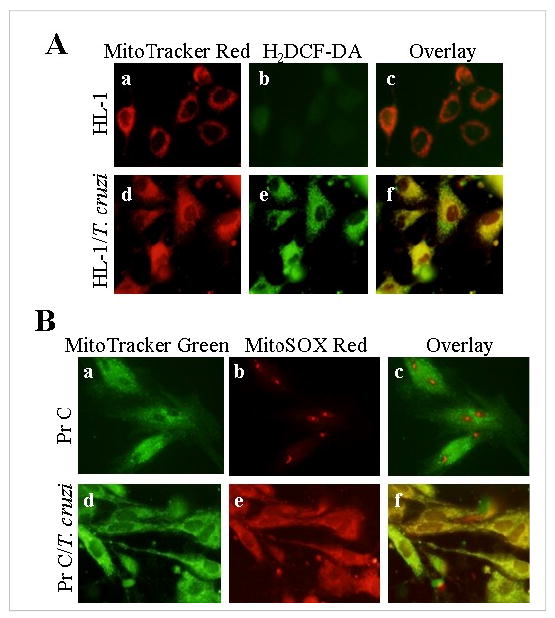
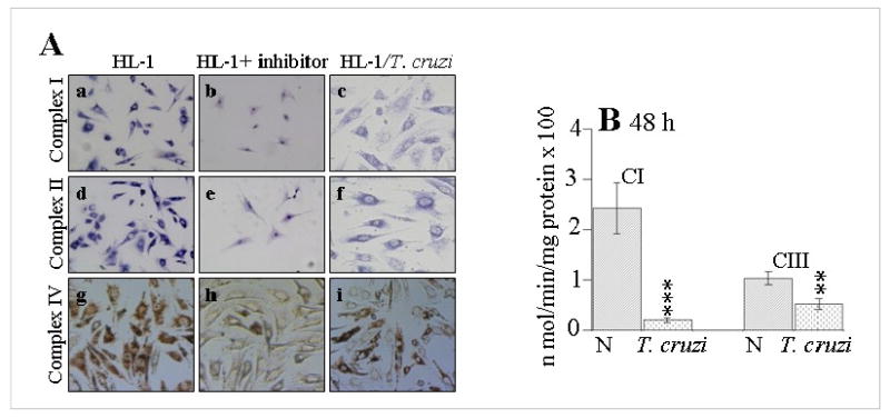
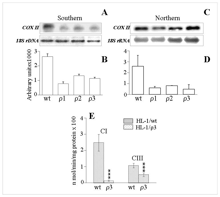
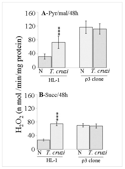
In this study, we investigated the role of Trypanosoma cruzi invasion and inflammatory processes in reactive oxygen species (ROS) production in a mouse atrial cardiomyocyte line (HL-1) and primary adult rat ventricular cardiomyocytes. Cardiomyocytes were incubated with T. cruzi (Tc) trypomastigotes, Tc lysate (TcTL), or Tc secreted proteins (TcSP) for 0-72 h, and ROS were measured by amplex red assay. Cardiomyocytes infected by T. cruzi (but not those incubated with TcTL or TcSP) exhibited a linear increase in ROS production for 2-48 h postinfection (max 18-fold increase), which was further enhanced by recombinant cytokines (IL-1beta, TNF-alpha, and IFN-gamma). We observed no increase in NADPH oxidase, xanthine oxidase, or myeloperoxidase activity, and specific inhibitors of these enzymes did not block the increased rate of ROS production in infected cardiomyocytes. Instead, the mitochondrial membrane potential was perturbed and resulted in inefficient electron transport chain (ETC) activity and enhanced electron leakage and ROS formation in infected cardiomyocytes. HL-1 rho (rho) cardiomyocytes lacked a functional ETC and exhibited no increase in ROS formation in response to T. cruzi. Together, these results demonstrate that invasion by T. cruzi and an inflammatory milieu affect mitochondrial integrity and contribute to electron transport chain inefficiency and ROS production in cardiomyocytes.


Figures

Figure 1
Figure 1. T. cruzi infection elicits ROS generation in cardiomyocytes
(A) HL-1 cardiomyocytes were infected with T. cruzi (cell: parasite ratio, 1:5), and ROS level in supernatant measured using amplex red fluorescent probe. (B) ROS generation in primary adult rat cardiomyocytes at 24 h and 48 h pi with T. cruzi. (C) HL-1 cardiomyocytes were infected with T. cruzi, or incubated with trypomastigote lysate (TcTL) or secreted proteins (TcSP) in presence or absence of catalase (CAT), and ROS level measured at 48 h pi. Data (mean ± SD) are representative of three independent experiments (**p <0.01, ***p <0.001, normal (N) versus T. cruzi-infected).
Figure 2
Figure 2. Proinflammatory cytokines augment the T. cruzi-induced ROS production in cardiomyocytes
(A) HL-1 cells were infected with T. cruzi, or incubated with TcTL or TcSP in presence or absence of 100 ng/ml each of IL-1β, TNF-α, and IFN-γ cytokines for 48 h and ROS level measured (as above). (B) Shown are H2O2 levels in primary cardiomyocytes infected with T. cruzi ± recombinant cytokines. Data (mean ± SD) are representative of triplicate observations (n = 3 experiments, **p < 0.01, normal versus infected). Significance (p < 0.05) of cytokine treatment is shown by dot-head.
Figure 3
Figure 3. Mitochondrial membrane potential (Δψ) is disturbed in cardiomyocytes infected by T. cruzi
(A & B) HL-1 and primary cardiomyocytes (PrC) were infected with T. cruzi, and at 48 h pi, loaded with JC-1 probe. Shown are the representative fluorescence micrographs of JC-1-stained HL-1 (A) and primary (B) cardiomyocytes. Note the accumulation of red J-aggregates in normal cardiomyocytes (panels a) and an accumulation of green monomers in infected cardiomyocytes (panels e). Overlay images show that a majority of mitochondria fluoresced red in normal cardiomyocytes (panels c) while mitochondria in infected cardiomyocytes fluoresced green (panels f). (C) ROS contributes to mitochondrial depolarization in T. cruzi-infected cardiomyocytes. HL-1 cardiomyocytes were infected with T. cruzi or incubated with 20 μM H2O2 in presence or absence of catalase (CAT). Shown are the ratios of fluorescence intensity of J-monomers (green fluorescence) to J-aggregates (red fluorescence). Data (mean ± SD) are representative of triplicate observations (n = 3 experiments, **p <0.01, normal versus H2O2-treated or T. cruzi-infected). Significance of catalase treatment (p < 0.05) is shown by dot-head.
Figure 4
Figure 4. Mitochondrial ROS release is increased in cardiomyocytes infected by T. cruzi
(A) Shown are the representative fluorescence micrographs of HL-1 normal (panels a, b, c) and T. cruzi-infected (panels d, e, f) cardiomyocytes at 48 h pi, stained with MitoTracker red (localizes to mitochondria, panels a, d) and H2DCFDA (detects intracellular ROS, green fluorescence, panels b, e). (B) Normal (panels a, b, c) and T. cruzi-infected (panels d, e, f) primary cardiomyocytes were stained at 48 h pi with MitoTracker green (localizes to mitochondria, panels a, d) and MitoSOX red (detects mitochondrial O2•−, panels b, e). Overlay images demonstrate co-localization of MitoTracker and ROS-specific probes in infected cardiomyocytes (panel f).
Figure 5
Figure 5. Electron transport chain activity is compromised in mitochondria of T. cruzi-infected cardiomyocytes
(A) Shown are representative histochemical micrographs for complex I (panels a, b & c), complex II (panels d, e & f) and complex IV (panels g, h & i) in normal HL-1 (panels a, d & g), HL-1 treated with specific inhibitors (b: rotenone, e: malonate, h: KCN), and infected HL-1 at 48 h pi (panels c, f & i). (B) Spectrophotometric evaluation of activities of complex I and complex III in mitochondria isolated from HL-1 cardiomyocytes at 48 h pi. Data (mean ± SD) are representative of triplicate observations (n = 3 experiments, (**p <0.01, ***p <0.001, normal versus infected).
Figure 6
Figure 6. HL-1 rho (ρ) cardiomyocytes are compromised in mitochondrial function
(A & B) Southern blotting. Total DNA from HL-1 wt and ρ cultures (ρ1, ρ2, ρ3) was digested with BamHI, and hybridization was performed with [32P]-labeled COX II or 18S rRNA probes. Densitometric analysis of COXII signal (indicative of mtDNA content), normalized with 18S signal (indicative of nuclear DNA) is shown in panel B. (C &D) Northern blot analysis. Total RNA was resolved on gel, and hybridized with [32P]-labeled cDNA probes for COX II or 18S rRNA. Densitometric analysis of COX II mRNA signal, normalized with 18S rRNA signal, is shown in panel D. (E) Mitochondria were isolated from HL-1 wt and ρ cardiomyocytes, and activities of complex I and complex III determined by spectrophotometry.
Figure 7
Figure 7. T. cruzi infection does not elicit ROS production in HL-1 ρ cardiomyocytes
HL-1 (wt and ρ3) cardiomyocytes were infected with T. cruzi. At 48 h pi, freshly isolated mitochondria were energized with pyruvate/malate (pyr/mal) (A) or succinate (B) substrates, and the rate of H2O2 production monitored using amplex red. Data (mean ± SD) are representative of triplicate observations (n = 3 experiments, **p <0.01, ***p <0.001, normal versus infected).

References

    1. World Health Organization. Report of the Scientific Working Group on Chagas Disease. UNDP/World Bank/WHO. 2006
    1. Higuchi MD. Endomyocardial biopsy in Chagas' heart disease: pathogenetic contributions. Sao Paulo Med J. 1995;113:821–825. - PubMed
    1. Zacks MA, Wen JJ, Vyatkina G, Bhatia V, Garg N. An overview of chagasic cardiomyopathy: pathogenic importance of oxidative stress. An Acad Bras Cienc. 2005;77:695–715. - PubMed
    1. Munoz-Fernandez MA, Fernandez MA, Fresno M. Activation of human macrophages for the killing of intracellular Trypanosoma cruzi by TNF-alpha and IFN-gamma through a nitric oxide-dependent mechanism. Immunol Lett. 1992;33:35–40. - PubMed
    1. Lima EC, Garcia I, Vicentelli MH, Vassalli P, Minoprio P. Evidence for a protective role of tumor necrosis factor in the acute phase of Trypanosoma cruzi infection in mice. Infect Immun. 1997;65:457–465. - PMC - PubMed

Publication types

MeSH terms