. 2009 Nov;77(11):5190-201.
doi: 10.1128/IAI.00420-09. Epub 2009 Aug 17.

Streptolysin S inhibits neutrophil recruitment during the early stages of Streptococcus pyogenes infection

Affiliations

Streptolysin S inhibits neutrophil recruitment during the early stages of Streptococcus pyogenes infection

Ada Lin et al. Infect Immun. 2009 Nov.

Abstract

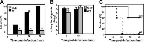
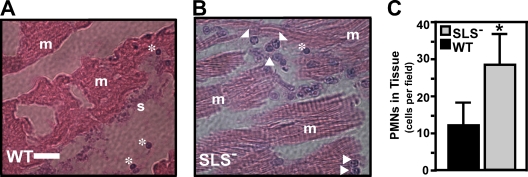
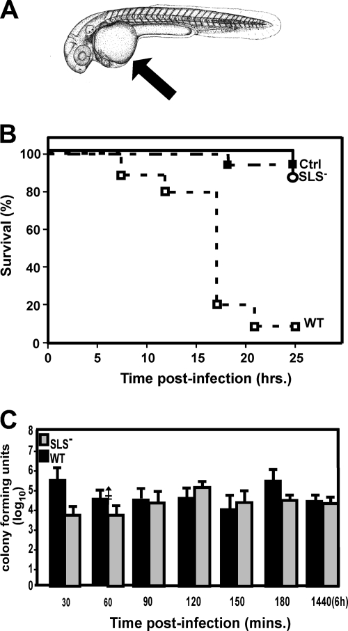
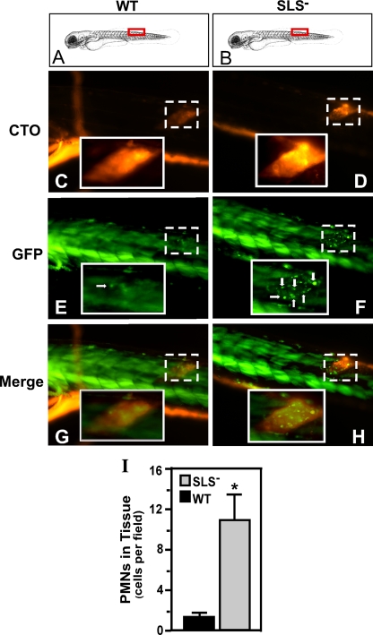
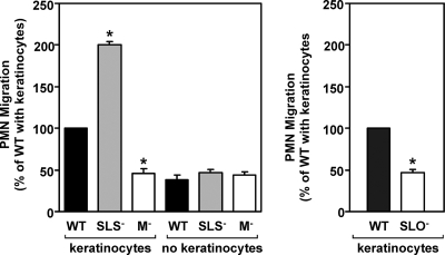
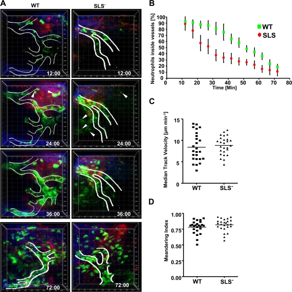
In contrast to infection of superficial tissues, Streptococcus pyogenes infection of deeper tissue can be associated with a significantly diminished inflammatory response, suggesting that this bacterium has the ability to both promote and suppress inflammation. To examine this, we analyzed the behavior of an S. pyogenes mutant deficient in expression of the cytolytic toxin streptolysin S (SLS-) and evaluated events that occur during the first few hours of infection by using several models including injection of zebrafish (adults, larvae, and embryos), a transepithelial polymorphonuclear leukocyte (PMN) migration assay, and two-photon microscopy of mice in vivo. In contrast to wild-type S. pyogenes, the SLS- mutant was associated with the robust recruitment of neutrophils and significantly reduced lethal myositis in adult zebrafish. Similarly, the mutant was attenuated in embryos in its ability to cause lethality. Infection of larva muscle allowed an analysis of inflammation in real time, which revealed that the mutant had recruited PMNs to the infection site. Analysis of transepithelial migration in vitro suggested that SLS inhibited the host cells' production of signals chemotactic for neutrophils, which contrasted with the proinflammatory effect of an unrelated cytolytic toxin, streptolysin O. Using two-photon microscopy of mice in vivo, we showed that the extravasation of neutrophils during infection with SLS- mutant bacteria was significantly accelerated compared to infection with wild-type S. pyogenes. Taken together, these data support a role for SLS in the inhibition of neutrophil recruitment during the early stages of S. pyogenes infection.


Figures

FIG. 1.
FIG. 1.
The SLS mutant is attenuated in the adult zebrafish model of necrotic myositis. Groups of zebrafish were challenged intramuscularly with 105 CFU of mutant (SLS) or wild-type (WT) S. pyogenes. Significant differences are indicated by an asterisk. (A) Percentage of fish with lesions at 6, 12, and 24 h. The data represent the average of 10 fish per experiment over three separate experiments at the indicated time points. There were significantly fewer lesions (P < 0.02) in the mutant group at each time point. (B) Numbers of CFU recovered from infected tissue at 6 and 12 h. Data represent the mean and standard deviation of three separate experiments. A significant difference was seen at 6 h but not at 12 h. (C) Survival data presented as a Kaplan-Meier plot. Data are pooled from three independent experiments, each of which was conducted using 10 zebrafish per group. These data indicate that the mutant was significantly less lethal than the wild type (P < 0.0043).
FIG. 2.
FIG. 2.
Infection by the SLS mutant demonstrates a neutrophil-rich infiltrate. Adult zebrafish were infected in the dorsal muscle by either the wild type (WT) or the SLS mutant, as indicated. Zebrafish from each infection group were fixed in toto. Longitudinal sections (5 μm thick) were stained with hematoxylin and eosin, and then the area of injection/infection was examined by microscopy. The panels show samples from 6 h postinfection with the wild-type strain HSC5 (A) and the SLS strain ΩSagH (B). Features apparent in the micrographs are denoted as follows: arrowheads, PMNs; asterisks, erythrocytes; m, striated muscle; and s, streptococci. The bar in the left panel represents 30 μm, and the center panel is shown at the same magnification. (C) Numbers of PMNs that infiltrated infected tissue as determined by microscopic examination of stained sections by blinded observers. Data represent the mean and standard deviation of the number of individual PMNs observed per microscopic field and are pooled from sections prepared from three infected zebrafish per experimental group. There were significantly more PMNs observed in tissue infected by the SLS mutant (P < 0.001).
FIG. 3.
FIG. 3.
S. pyogenes is virulent in zebrafish embryos but, as in adult zebrafish, the SLS mutant is not as virulent. (A) Yolk sac injections. The site of injection of 1 × 10⁵ CFU of wild-type (WT) S. pyogenes, the SLS mutant, or PBS alone (Ctrl) in the yolk sac is indicated by the arrow. The diagram was adapted from Kimmel et al. (39) with permission of the publisher, John Wiley & Sons, Inc. (B) Analysis of virulence. The survival data of injected embryos are presented as a Kaplan-Meier plot. Infected embryos were examined for viability every 2 h by microscopy. Data shown are pooled from three independent experiments using 15 embryos per group. These data show that the SLS mutant is significantly attenuated (P < 0.011). (C) Infected embryos were sacrificed every 30 min during the first 6 h of infection for assessment of the number of CFU. Groups of five embryos each were examined at each time point for each strain analyzed. The data shown represent the mean and standard deviation of three independent experiments. There was no significant difference between groups.
FIG. 4.
FIG. 4.
The SLS mutant also promotes a PMN-rich infiltrate in embryos. pu.1 larvae (7 days postfertilization) which express GFP fluorescence specifically in cells of the myeloid lineage were injected in the dorsal muscle with 103 CFU of either the wild type (WT) or the SLS mutant intrinsically labeled with the fluorescent dye CTO at the site indicated by the red boxes in panels A and B. Infected larvae were examined by fluorescent microscopy at 2 h postinfection for CTO fluorescence (C and D and GFP fluorescence (E and F; PMNs are indicated by white arrows). As noted by Hsu et al., in the larval form, the skeletal muscle (even in wild-type-infected fish) contributes background fluorescence that is easily distinguished from individual myeloid cells (36). The columns show images taken from single representative larvae, oriented as indicated in the top panels (A and B), that were infected with the wild type (WT) or the SLS mutant. Panels G and H show the merged images. The site of injection in each fish is demarcated in the micrographs by the boxes with the broken white line. Magnification, ×10. The insets outlined by the boxes with the unbroken white lines show the injection site at a higher magnification (×40). The diagram in panels A and B was adapted from Kimmel et al. (39) with permission of the publisher, John Wiley & Sons, Inc. (I) The number of PMNs that infiltrated the infected tissue as determined by microscopic examination by blinded observers. Data shown represent the mean and standard deviation of the number of individual PMNs observed per microscopic field and pooled from three infected zebrafish per experimental group. As with the results of infections of adults, there were significantly more PMNs observed in tissue infected by the SLS mutant than in wild-type-infected tissue (P < 0.0001).
FIG. 5.
FIG. 5.
Neutrophil recruitment is altered by SLS and requires the presence of keratinocytes. The number of PMNs recruited to the basolateral surface of a polarized HaCaT keratinocyte monolayer (keratinoctyes) following infection with various S. pyogenes strains. Monolayers were infected at their apical surfaces at 10 CFU/epithelial cell, and then 1 × 10⁶ PMNs were added at the basolateral surface of the monolayer. The number of PMNs recruited to the apical surface was enumerated at 60 min postinfection and is shown relative to the number recruited in response to each wild-type strain. PMN recruitment in the absence of a monolayer is also shown (no keratinoctyes). In the left panel, strains derived from HSC5 (WT) include the SLS mutant ΩSagH and the M protein-deficient mutant ΩEmm (M). In the right panel, the SLO-deficient mutant SLO1 (SLO) was derived from JRS4 (WT). An asterisk indicates that recruitment by the indicated strain was significantly different from the wild type (P < 0.001) under the same conditions. Data shown represent the mean and standard deviation of the mean derived from three independent experiments.
FIG. 6.
FIG. 6.
Characterization of PMN exposed to S. pyogenes in vitro under conditions of transwell infections. (A) The ability of PMNs to produce a respiratory burst in the presence of keratinocytes infected with wild-type (WT) or SLS S. pyogenes is shown. Human PMNs loaded with an indicator dye that fluoresces in the presence of intracellular ROS were tested in the transepithelial migration assays, and the data are presented as the amount of fluorescence (relative fluorescent units [RFU]) per 104 migrated cells. Data represent the mean and standard deviation of six samples from two independent experiments. (B) The number of PMNs undergoing apoptosis when exposed to wild-type (WT) or SLS S. pyogenes is shown, as determined by staining with annexin V-PE. Data presented show the ratio of the number of apoptotic cells observed upon exposure to the indicated strain versus the number observed in the absence of any streptococci and represent the mean and standard deviation of two independent experiments, with samples analyzed in duplicate. Differences between mean values at each time point shown in both panels were not significant.
FIG. 7.
FIG. 7.
In vivo imaging of neutrophil recruitment in mice infected with wild-type (WT) and SLS streptococci. Intravital imaging was performed at the injection site on the paws of LysM-eGFP mice after subcutaneous infection with streptococci. Data were pooled from three independent experiments. (A) Representative time-lapse images of neutrophil recruitment in response to wild-type and SLS mutant bacteria. Images are three-dimensionally rendered volumes (200 μm by 225 μm by 75 μm). Neutrophils (eGFP) appear green, collagen fibers in the connective tissue appear blue, and blood vessels (unless occluded by cells) appear red. White lines show the outline of blood vessels based on both the fluorescent-dextran signal and the characteristic flow of circulating neutrophils. White arrowheads show extravasated neutrophils at 24 min. The time stamp indicates minutes postinfection. The scale grid is labeled in micrometers. (B) The percentage ± standard deviation of neutrophils remaining in blood vessels over time in LysM-GFP mice infected with wild-type and SLS bacteria. The curves are statistically different (P < 0.01) with both Pearson (parametric) and Spearman (nonparametric) correlations. (C) Interstitial neutrophil migration velocity. The plot shows the median track velocities of 25 extravasated neutrophils in wild-type and SLS mutant infections. No statistical differences were observed. (D) The meandering index was calculated for each track in panel C. The median meandering indices of neutrophils after wild-type and SLS mutant infection were not statistically different.
FIG. 8.
FIG. 8.
IL-8 expression is similar in transwells infected with either wild-type or SLS-deficient streptococci. Polarized HaCaT keratinocyte monolayers were treated with 1 × 10⁶ bacteria/monolayer of wild-type (WT) bacteria, an equivalent concentration of SLS bacteria, or an equivalent volume of chemotaxis medium alone as a control (Ctrl). The monolayers were assessed for IL-8 secretion in the apical and basolateral compartments. Each strain was represented by 10 wells per experiment, and data presented are representative of an experiment performed three times.

References

    1. Bakleh, M., L. E. Wold, J. N. Mandrekar, W. S. Harmsen, H. H. Dimashkieh, and L. M. Baddour. 2005. Correlation of histopathologic findings with clinical outcome in necrotizing fasciitis. Clin. Infect. Dis. 40:410-414. - PubMed
    1. Bernheimer, A. W. 1967. Physical behavior of streptolysin S. J. Bacteriol. 93:2024-2025. - PMC - PubMed
    1. Betschel, S. D., S. M. Borgia, N. L. Barg, D. E. Low, and J. C. De Azavedo. 1998. Reduced virulence of group A streptococcal Tn916 mutants that do not produce streptolysin S. Infect. Immun. 66:1671-1679. - PMC - PubMed
    1. Boukamp, P., R. T. Petrussevska, D. Breitkreutz, J. Hornun, A. Markham, and N. E. Fusenig. 1988. Normal keratinization in a spontaneously immortalized aneuploid human keratinocyte one. J. Cell Biol. 106. - PMC - PubMed
    1. Caparon, M. G., D. S. Stephens, A. Olsen, and J. R. Scott. 1991. Role of M. protein in adherence of group A streptococci. Infect. Immun. 59:1811-1817. - PMC - PubMed

Publication types

MeSH terms