. 2009 Sep;134(3):231-41; S1-2.
doi: 10.1085/jgp.200910272. Epub 2009 Aug 17.

Different pathways for activation and deactivation in CaV1.2: a minimal gating model

Affiliations

Different pathways for activation and deactivation in CaV1.2: a minimal gating model

Stanislav Beyl et al. J Gen Physiol. 2009 Sep.

Abstract

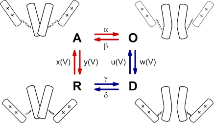
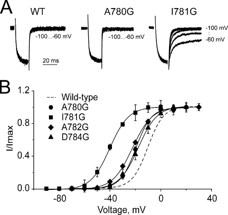
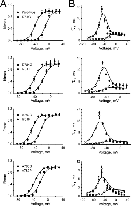
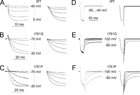
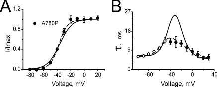
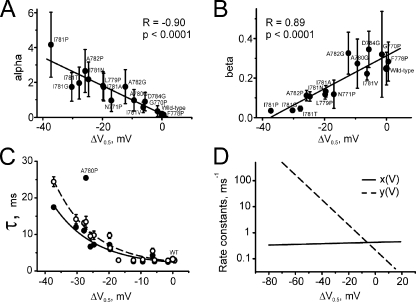
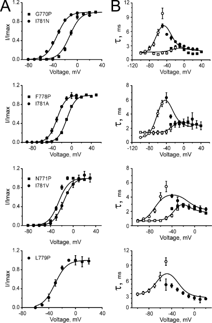
Point mutations in pore-lining S6 segments of CaV1.2 shift the voltage dependence of activation into the hyperpolarizing direction and significantly decelerate current activation and deactivation. Here, we analyze theses changes in channel gating in terms of a circular four-state model accounting for an activation R-A-O and a deactivation O-D-R pathway. Transitions between resting-closed (R) and activated-closed (A) states (rate constants x(V) and y(V)) and open (O) and deactivated-open (D) states (u(V) and w(V)) describe voltage-dependent sensor movements. Voltage-independent pore openings and closures during activation (A-O) and deactivation (D-R) are described by rate constants alpha and beta, and gamma and delta, respectively. Rate constants were determined for 16-channel constructs assuming that pore mutations in IIS6 do not affect the activating transition of the voltage-sensing machinery (x(V) and y(V)). Estimated model parameters of 15 CaV1.2 constructs well describe the activation and deactivation processes. Voltage dependence of the "pore-releasing" sensor movement ((x(V)) was much weaker than the voltage dependence of "pore-locking" sensor movement (y(V)). Our data suggest that changes in membrane voltage are more efficient in closing than in opening CaV1.2. The model failed to reproduce current kinetics of mutation A780P that was, however, accurately fitted with individually adjusted x(V) and y(V). We speculate that structural changes induced by a proline substitution in this position may disturb the voltage-sensing domain.


Figures

Figure 1.
Figure 1.
Schematic representation of CaV1.2 state transitions during activation. Activation gating assumed to be determined by two functionally separate processes: a voltage-sensing mechanism (++) and the conducting pore. Each functional unit can dwell in two states: the voltage sensor in the resting (down) and activated (up) states, and the pore in the open or closed states. The entire molecule therefore dwells in 2 × 2 = 4 states: R, pore is closed and voltage-sensing mechanism locks the pore; A, voltage-sensing mechanism is activated and releases the pore, which, however, remains closed; O, the pore is open; D, the deactivated voltage-sensing mechanism is in the down position while the pore is still open. The activation pathway is marked by red and the deactivation pathway by blue arrows. Rate constants of the pore opening and closure (α, β, γ, and δ) are assumed to be independent of the voltage. Rate constants of voltage-sensing mechanism (x, y, u, and w) are voltage dependent.
Figure 2.
Figure 2.
Glycine mutations in IIS6 differentially modulate CaV1.2 gating. (A) Representative tail currents of wild-type, A780G, and I781G channels. Currents were activated during a 20-ms conditioning depolarization to 0 mV for wild-type, −10 mV for A780P, and −30 mV for I781G. Deactivation was recorded during subsequent repolarizations (10-mV increments) starting from −100 mV. (B) Voltage dependences of steady-state activation of the indicated glycine mutants.
Figure 3.
Figure 3.
Effects of pore mutations on steady-state activation and current kinetics. Voltage dependences of steady-state activation (A), time constants of activation (B; filled symbols), and deactivation (B; open symbols) of the indicated channel construct. Solid lines represent model simulations based on the identified model parameters given in Tables II and III.
Figure 4.
Figure 4.
Recorded and simulated currents. (A–C) IBa through wild-type (A), I781G (B), and I781P (C) channels (left column) during depolarization from −80 mV to the indicated voltages and corresponding current simulations (right column). (D–F) Representative tail currents of wild-type (D), I781G (E), and I781P (F) channels (left column) and corresponding simulations (right column). The fast phase of the tail current (D–F) containing fast (not resolved) components of current deactivation is truncated. The simulated currents were calculated from the full four-state model by solving the ODE system (Eq. 4.5) making use of the estimated rate constants (Tables II and III) and a reversal potential of 48 mV (see also Materials and methods).
Figure 5.
Figure 5.
Steady-state activation and current kinetics of A780P. Voltage dependence of steady-state activation (A) and time constants of activation and deactivation (B) of mutant A780P. Solid lines show simulations based on the rate constants x(V) and y(V) of Table III. Voltage dependence of the activation time constants (B) was not reproduced under the assumption of x(V) and y(V) being identical for all mutants. Dotted lines show improved simulations for A780P obtained by individual fits of x(V) and y(V) (see Table IV).
Figure 6.
Figure 6.
Rate constants of channel opening and closure. (A and B) Correlations between estimated rate constants of opening (A) and closure (B) and shifts of the activation curves (ΔV = V1/2,MUT − V1/2,WT) for IIS6 mutations. (C) Maximal time constants experimentally measured (open circles) and calculated (filled circles; see bell-shaped curves in Figs. 3 and 8) versus corresponding shifts of the activation curves. Lines represent fits by monoexponential functions (A0 + A1 * exp(−ΔV/k)), with A0 = 1.57 ± 1.10, A1 = 1.09 ± 0.52, and k =12.2 ± 1.9 for experimental data, and A0 = 1.53 ± 0.95, A1 = 0.88 ± 0.49, and k = 12.7 ± 2.3 for calculated data. (D) Voltage dependence of x(V) and y(V) plotted in semilogarithmic scale. x(V) represents the voltage dependence of the forward rate and y(V) the backward movement of the voltage-sensing machinery (see Fig. 1). A steeper voltage dependence of y(V) is evident.
Figure 7.
Figure 7.
Voltage dependence of deactivation time constants predicted by fitting the data for mutant I781T to the backward activation pathway: OAR. Deactivation time constant (solid line) was calculated by Eq. 2.2 (rate constants are indicated in Tables II and III). The model predicts an increase of the deactivation time constants, whereas the experimentally measured time constants decreased with increasing hyperpolarization (open circles connected by broken line).
Figure 8.
Figure 8.
Steady-state and kinetic characteristics of previously described mutants (Hohaus et al., 2005). Voltage dependencies of steady-state activation (A), time constants of activation (B; filled symbols), and deactivation (B; open symbols) of the indicated constructs. Solid lines represent the simulations based on the model parameters identified (see Tables II and III).

References

    1. Barlow D.J., Thornton J.M. 1988. Helix geometry in proteins.J. Mol. Biol. 201:601–619 - PubMed
    1. Bates D.M., Watts D.G. 1988. Nonlinear Regression Analysis and Its Application. Wiley Series in Probability and Mathematical Statistics, Inc. John Wiley and Sons, New York: 384 pp
    1. Catterall W.A. 2000. Structure and regulation of voltage-gated Ca2+ channels.Annu. Rev. Cell Dev. Biol. 16:521–555 - PubMed
    1. Cordes F.S., Bright J.N., Sansom M.S. 2002. Proline-induced distortions of transmembrane helices.J. Mol. Biol. 323:951–960 - PubMed
    1. Doyle D.A., Morais Cabral J., Pfuetzner R.A., Kuo A., Gulbis J.M., Cohen S.L., Chait B.T., MacKinnon R. 1998. The structure of the potassium channel: molecular basis of K+ conduction and selectivity.Science. 280:69–77 - PubMed

Publication types