. 2009 Aug 24;186(4):615-28.
doi: 10.1083/jcb.200903102. Epub 2009 Aug 17.

Steric and not structure-specific factors dictate the endocytic mechanism of glycosylphosphatidylinositol-anchored proteins

Affiliations

Steric and not structure-specific factors dictate the endocytic mechanism of glycosylphosphatidylinositol-anchored proteins

Pinkesh Bhagatji et al. J Cell Biol. .

Abstract

Diverse glycosylphosphatidylinositol (GPI)-anchored proteins enter mammalian cells via the clathrin- and dynamin-independent, Arf1-regulated GPI-enriched early endosomal compartment/clathrin-independent carrier endocytic pathway. To characterize the determinants of GPI protein targeting to this pathway, we have used fluorescence microscopic analyses to compare the internalization of artificial lipid-anchored proteins, endogenous membrane proteins, and membrane lipid markers in Chinese hamster ovary cells. Soluble proteins, anchored to cell-inserted saturated or unsaturated phosphatidylethanolamine (PE)-polyethyleneglycols (PEGs), closely resemble the GPI-anchored folate receptor but differ markedly from the transferrin receptor, membrane lipid markers, and even protein-free PE-PEGs, both in their distribution in peripheral endocytic vesicles and in the manner in which their endocytic uptake responds to manipulations of cellular Arf1 or dynamin activity. These findings suggest that the distinctive endocytic targeting of GPI proteins requires neither biospecific recognition of their GPI anchors nor affinity for ordered-lipid microdomains but is determined by a more fundamental property, the steric bulk of the lipid-anchored protein.


Figures
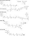
Figure 1.
Figure 1.
Structures of PE-PEG ligand conjugates and fluorescent lipid markers. Structures of the PE-PEG ligand conjugates used in this study (R1 and R2 are alkyl chains with varying structures, as indicated in the text; asterisks indicate sites of linkage of group X to the PE-PEG anchor), the bulk lipid marker DLMal-TMR, and the fluorescent PE-PEG species DL1500-TMR are shown. Anti-DNP antibody binds strongly to the TNP group of PE-PEG-TNP but can be displaced at acidic pH in the presence of the competing ligand ϵ–DNP-lysine. Streptavidin binds tightly to the biotinyl residue of PE-PEGSS-biotin but can be released by cleavage of the disulfide bond (arrow) in thiol-containing media. E. coli DHFR binds tightly to PE-PEG-methotrexate but can be displaced by free methotrexate at neutral pH.
Figure 2.
Figure 2.
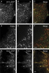
Internalized anti-DNP antibody bound to a di16:0PEG anchor colocalizes with internalized transferrin in a central endosomal compartment but not in the cell periphery. Cells pretreated to incorporate di16:0PEG-TNP were incubated for 10 min at 37°C with Alexa Fluor 488–labeled anti-DNP antibody and Alexa Fluor 555–transferrin and then rapidly chilled, stripped of surface-bound labels, and fixed before microscopic examination as described in Materials and methods. (A–C) Confocal images of the distribution of transferrin (A) and lipid-anchored anti-DNP antibody (B; C shows the merged image) show a strong coincidence of fluorescence labeling in the central region of the cell. The images shown in A and B were acquired under conditions in which the fluorescence intensity in the central region of the cell was not saturated and in which the peripheral region accordingly shows only weak fluorescence. (D) Wide-field microscopic image (with central fluorescence shown at saturating levels to reveal peripheral structures) illustrating the divergent distributions of lipid-anchored antibody (green) and transferrin (red) at the cell periphery. Bars, 10 µm.
Figure 3.
Figure 3.
Anti-DNP antibody bound to a di16:0PEG lipid anchor is internalized into peripheral endocytic structures that are largely distinct from transferrin-containing vesicles but that contain the GPI-linked folate receptor. Cells pretreated to incorporate di16:0PE-PEG-TNP were incubated with the indicated pairs of endocytic markers for 10 min at 37°C and then rapidly chilled, stripped of surface-bound fluorescent labels, and fixed before microscopic observation as described in Materials and methods. (A–C) Cells coincubated with Alexa Fluor 488–anti-DNP antibody (green in merged image) and Alexa Fluor 555–transferrin (Tf). (D–F) Cells coincubated with fluorescein-labeled folate (green in merged image) and Alexa Fluor 555–transferrin. (G–I) Cells coincubated with Alexa Fluor 488–anti-DNP antibody (green in merged image) and rhodamine-labeled folate. Bars, 2 µm.
Figure 4.
Figure 4.
Quantitative analysis of colocalization of di16:0PE-PEG–anchored proteins with transferrin and folate in endocytic vesicles. (A) Cells incorporating di16:0PE-PEG-TNP or di16:0PE-PEGSS-biotin were coincubated with the indicated pairs of fluorescent markers (SAv, streptavidin; Tf, transferrin) for 10 min at 37°C, and then the extent of colocalization of the two markers in peripheral endocytic structures was determined by microscopy after removal of surface-bound markers and cell fixation (for further details, see Materials and methods). Data labeled folate/folate and Tf/Tf represent the values of the co-distribution index in peripheral endocytic vesicles measured for cells coincubated with fluorescein- and rhodamine-labeled folate or with Alexa Fluor 488– and Alexa Fluor 555–labeled transferrin, respectively, and indicate the value of the co-distribution index measured for species internalized in an identical manner. (B and C) Cells incorporating PE-PEG-TNP, PE-PEGSS-biotin or PE-PEG-methotrexate species with the indicated acyl or alkyl chains were incubated with labeled anti-DNP antibody, streptavidin, or DHFR, as appropriate, together with labeled transferrin (B) or folate (C), and the extent of colocalization of the two markers in peripheral vesicles was determined as described for A. Values plotted in A–C represent the mean (±SEM) of determinations for 30–60 separate fields (all representing distinct cells) in three to five independent experiments.
Figure 5.
Figure 5.
di16:0PE-PEG–anchored streptavidin colocalizes only weakly with transferrin but strongly with the GPI-linked folate receptor in peripheral endocytic vesicles. (A–F) Cells incorporating di16:0PE-PEGSS-biotin (structure shown in Fig. 1) were allowed to endocytose Alexa Fluor 555–labeled streptavidin (SAv; red in merged images) along with either Alexa Fluor 488–labeled transferrin (Tf; A–C) or fluorescein-labeled folate (D–F) for 10 min at 37°C and then rapidly chilled, stripped of surface-bound fluorescent labels, and fixed before microscopic observation. Other experimental details were as described in Materials and methods. Bars, 2 µm.
Figure 6.
Figure 6.
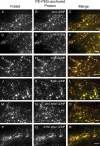
Proteins anchored to PE-PEG anchors with different hydrocarbon chain structures colocalize with the GPI-anchored folate receptor in peripheral endocytic vesicles. (A–R) Cells incorporating the indicated diacyl/dialkyl–PE-PEG-TNP or –PE-PEG-biotinyl anchors were incubated for 10 min with Alexa Fluor 555–anti-DNP antibody or Alex Fluor 555–streptavidin together with fluorescein-labeled folate (green in merged images) and then chilled, stripped of surface-bound fluorescent molecules, and fixed for fluorescence imaging. Anchor species and bound fluorescent proteins are designated as follows: di12-, di16c-, and d18c–α-DNP indicate anti-DNP antibody bound to di12:0-, di16:1c-, or di18:1cPE-PEG-TNP; di12-SAv indicates streptavidin bound to di12:0PE-PEGSS-biotin; di16 ether- and di18c ether–α-DNP indicate anti-DNP antibody bound to dihexadecyl– or di-cis-9′-octadecenyl–PE-PEG-TNP. Other experimental details were as described in Materials and methods. Bars, 2 µm.
Figure 7.
Figure 7.
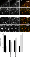
A bulk lipid marker and a fluorescent PE-PEG label both transferrin- and folate-containing peripheral endocytic vesicles. Cells were preincubated for 30 min at 4°C to incorporate DLMal-TMR or the fluorescent PE-PEG DL1500-TMR (structures shown in Fig. 1), fluorescent transferrin or folate was added, and the cells were rapidly warmed to 37°C. After 10 min, the cells were chilled, residual surface-bound fluorescent markers were removed, and the fixed cells were imaged by fluorescence microscopy. (A–F) Cells coincubated at 37°C with DLMal-TMR (lipid; red in merged images) and either Alexa Fluor 488–transferrin (Tf) or fluorescein-labeled folate. (G–L) Cells coincubated at 37°C with DL1500-TMR (PEPEG; red in merged images) and either Alexa Fluor 488–transferrin or fluorescein-labeled folate. The virtual absence of deep green vesicles (labeled by transferrin or folate only) illustrates the observation that essentially all transferrin- and folate-containing vesicles are also labeled to some extent by the lipid and PE-PEG markers. (M) Quantitation of the indexes of co-distribution of DLMal-TMR and DL1500-TMR with transferrin (solid bars) and folate (hatched bars). Values plotted represent the mean (±SEM) of determinations for 40–50 separate fields (all from distinct cells) in three to four independent experiments. The co-distribution indexes measured in parallel experiments for cointernalized Alexa Fluor 488– and Alexa Fluor 555–transferrin (Tf solid bar), fluorescein- and rhodamine-labeled folate (Folate hatched bar), and labeled folate and transferrin (Tf hatched bar and Folate solid bar) are shown for reference. Other experimental details are described in Materials and methods. Bars, 2 µm.
Figure 8.
Figure 8.
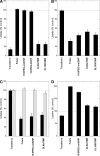
Modulation of dynamin and Arf1 activities affects endocytosis of artificially lipid-anchored proteins in a manner very similar to that observed for GPI proteins. Cells were allowed to internalize Alexa Fluor 555–transferrin, rhodamine-labeled folate, Alexa Fluor 555–labeled anti-DNP antibody (for cells incorporating di16:0PE-PEG-TNP), or DHFR (for cells incorporating di16:0PE-PEG-methotrexate), the bulk lipid marker DLMal-TMR, or the fluorescent PE-PEG DL1500-TMR (the latter two markers were preincorporated into the plasma membrane at 0°C) for 3 min at 37°C. The amount of internalized marker was then determined by microscopy after the removal of residual surface-associated markers and fixation as described in Materials and methods. Uptake of each fluorescence marker under the indicated treatments is expressed as a percentage of the level of marker internalization measured in parallel untreated control samples. Data values shown represent the mean (±SEM) determined from three to seven independent experiments. (A) Effects of 80 µM of the dynamin inhibitor dynasore preincubated with cells for 20 min. (B) Effects of transient expression of a dominant-negative (T31N) form of Arf1. (C) Effects of expression of shRNAs directed against Arf1 (solid bars) or against Arf3 as a negative control (hatched bars). (D) Effects of transient expression of a constitutively activated (Q71L) form of Arf1.

Comment in

References

    1. Barlos K., Gatos D., Hatzi O., Koch N., Koutsogianni S. 1996. Synthesis of the very acid-sensitive Fmoc-Cys(Mmt)-OH and its application in solid-phase peptide synthesis.Int. J. Pept. Protein Res. 47:148–153 - PubMed
    1. Barr D.J., Ostermeyer-Fay A.G., Matundan R.A., Brown D.A. 2008. Clathrin-independent endocytosis of ErbB2 in geldanamycin-treated human breast cancer cells.J. Cell Sci. 121:3155–3166 - PMC - PubMed
    1. Bonazzi M., Spanò S., Turacchio G., Cericola C., Valente C., Colanzi A., Kweon H.S., Hsu V.W., Polishchuck E.V., Polishchuck R.S., et al. 2005. CtBP3/BARS drives membrane fission in dynamin-independent transport pathways.Nat. Cell Biol. 7:570–580 - PubMed
    1. Chadda R., Howes M.T., Plowman S.J., Hancock J.F., Parton R.G., Mayor S. 2007. Cholesterol-sensitive Cdc42 activation regulates actin polymerization for endocytosis via the GEEC pathway.Traffic. 8:702–717 - PMC - PubMed
    1. Cheng Z.J., Singh R.D., Sharma D.K., Holicky E.L., Hanada K., Marks D.L., Pagano R.E. 2006. Distinct mechanisms of clathrin-independent endocytosis have unique sphingolipid requirements.Mol. Biol. Cell. 17:3197–3210 - PMC - PubMed

Publication types

MeSH terms