. 2009 Aug 24;186(4):541-54.
doi: 10.1083/jcb.200904050. Epub 2009 Aug 17.

Myosin-driven peroxisome partitioning in S. cerevisiae

Affiliations

Myosin-driven peroxisome partitioning in S. cerevisiae

Andrei Fagarasanu et al. J Cell Biol. .

Abstract

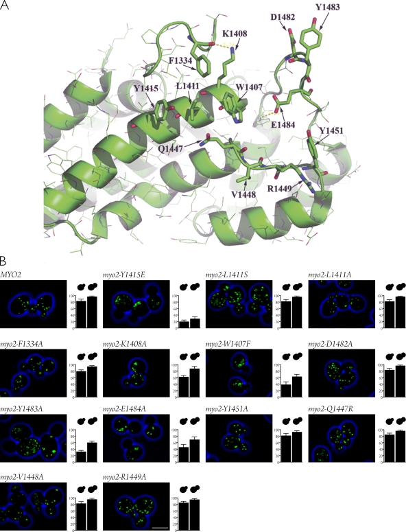
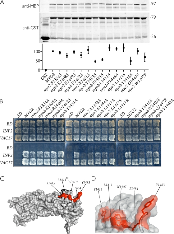
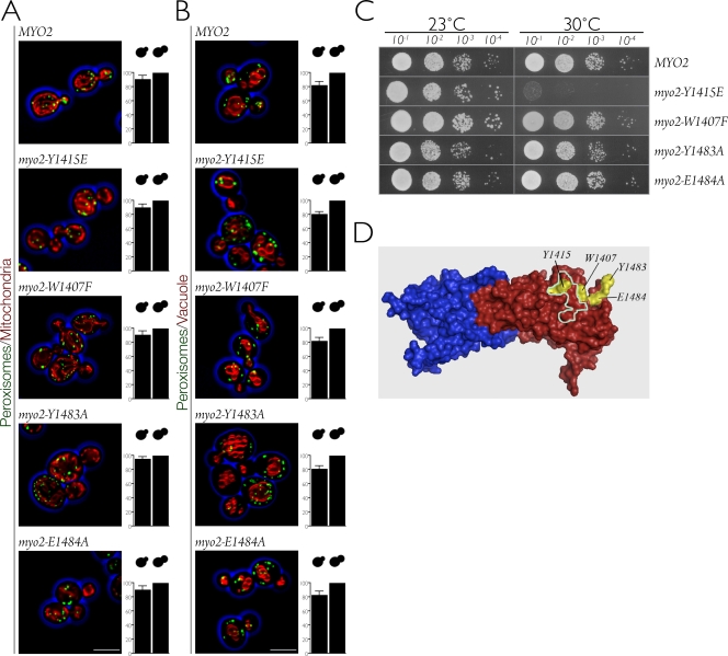
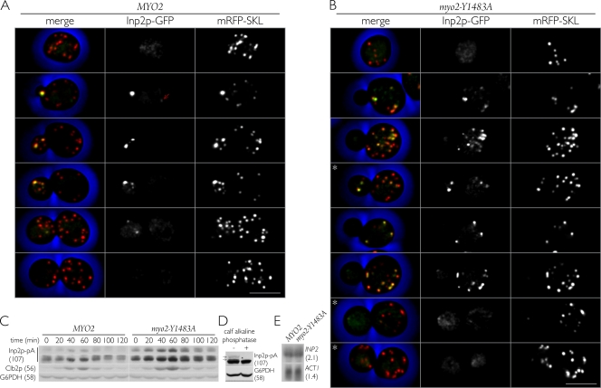
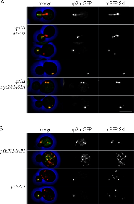
In Saccharomyces cerevisiae, the class V myosin motor Myo2p propels the movement of most organelles. We recently identified Inp2p as the peroxisome-specific receptor for Myo2p. In this study, we delineate the region of Myo2p devoted to binding peroxisomes. Using mutants of Myo2p specifically impaired in peroxisome binding, we dissect cell cycle-dependent and peroxisome partitioning-dependent mechanisms of Inp2p regulation. We find that although total Inp2p levels oscillate with the cell cycle, Inp2p levels on individual peroxisomes are controlled by peroxisome inheritance, as Inp2p aberrantly accumulates and decorates all peroxisomes in mother cells when peroxisome partitioning is abolished. We also find that Inp2p is a phosphoprotein whose level of phosphorylation is coupled to the cell cycle irrespective of peroxisome positioning in the cell. Our findings demonstrate that both organelle positioning and cell cycle progression control the levels of organelle-specific receptors for molecular motors to ultimately achieve an equidistribution of compartments between mother and daughter cells.


Figures

Figure 1.
Figure 1.
Surface representation of the Myo2p globular tail. Subdomains I and II are shown in blue and red, respectively. The mutated, conserved surface residues initially screened for defects in peroxisome inheritance are shown in white. The yellow outline demarcates the vacuole-binding site, and the teal outline demarcates the secretory vesicle–binding site.
Figure 2.
Figure 2.
Screening mutations in Myo2p for defects in peroxisome inheritance. (A) Ribbon diagram of a portion of subdomain II of Myo2p highlighting the amino acids that were mutated. (B) Quantification of peroxisome inheritance. The percentages of buds containing peroxisomes at each size category were plotted. Quantification was performed on at least 50 budded cells from each category. Graphic results show the means ± SEM of three independent experiments. Bar, 5 µm.
Figure 3.
Figure 3.
Mutants of Myo2p defective in peroxisome inheritance display decreased affinity for Inp2p. (A) Glutathione Sepharose beads containing either GST fused to the cargo-binding tail of wild-type or mutant Myo2 proteins or GST alone were incubated with extracts of E. coli–synthesized MBP–Inp2p. (top) Bound MBP–Inp2p was analyzed by immunoblotting with anti-MBP antibodies. (middle) Total GST–Myo2p or GST protein levels were visualized by immunoblotting with anti-GST antibodies. Numbers at right denote the approximate sizes of proteins in kilodaltons. (bottom) Quantification of bound MBP–Inp2p, normalized to wild type and expressed as percent bound, was performed by densitometry. Graphic results show the means ± SEM of three independent experiments. (B) Myo2p point mutations specifically disrupt the ability of Myo2p to interact with Inp2p (peroxisomes) but not with Vac17p (vacuoles) in a yeast two-hybrid assay. Total growth of strains (top) and growth arising specifically from protein interaction (bottom) are shown. The pattern presented is representative of seven independent experiments. AD, activation domain; BD, binding domain. (C) Crystal structure of the Myo2p globular tail with the positions of point mutations that showed either weak or no interaction with Inp2p highlighted in red. The asterisk at L1411 indicates that the L1411R mutation was able to disrupt the Myo2p–Inp2p interaction in the yeast two-hybrid assay. The secretory vesicle–binding region is outlined in black. (D) Semitransparent surface view of the Myo2p structure overlaid on a ribbon diagram displaying the side chains of residues (red) implicated in binding Inp2p.
Figure 4.
Figure 4.
Myo2p point mutants defective in peroxisome inheritance are not impaired in vacuolar or mitochondrial inheritance. Cells expressing wild-type MYO2 or mutant myo2 were grown in SCIM to induce peroxisome formation, and confocal images were captured. (A and B) Peroxisomes were labeled with Pot1p-GFP, mitochondria were labeled with MitoTracker red (A), and vacuoles were labeled with FM4-64 (B). Quantification of mitochondria and vacuole inheritance was performed as in Fig. 2 B. Graphic results show the means ± SEM of three independent experiments. Bars, 5 µm. (C) Growth of cells expressing wild-type MYO2 or mutant myo2 on glucose-containing YPD medium. Cells were grown to mid-log phase in liquid YPD, and equal amounts of cells were serially diluted 10-fold onto YPD agar and incubated at 23 or 30°C. (D) Surface representation of the Myo2p globular tail, indicating the regions that bind peroxisomes (yellow) and secretory vesicles (teal). Subdomains I and II are shown in blue and red, respectively.
Figure 5.
Figure 5.
Dynamics of Inp2p in cells defective in peroxisome transfer to buds. (A and B) Inp2p accumulates on most peroxisomes in mother cells of myo2 mutants defective in peroxisome inheritance. Cells harboring the MYO2 gene (A) or myo2-Y1483A mutation (B) and expressing Inp2p-GFP and peroxisomal mRFP-SKL were grown in YPD to mid-log phase, and confocal images were captured. Left panels show the merged image of the middle and right panels. The red arrow indicates a peroxisome in a mother cell of the MYO2 strain that contains a detectable amount of Inp2p-GFP. Asterisks indicate myo2-Y1483A mutant cells that have delivered peroxisomes to their buds. Bars, 5 µm. (C) Inp2p levels are increased in cells defective in peroxisome inheritance. Cells harboring either MYO2 or the myo2-Y1483A mutant allele and expressing Inp2p-pA were grown in YPD and synchronized in G1 by addition of α factor (0 min). After removal of α factor, cells were incubated at 23°C in YPD. Samples were collected at the times indicated, and total cell lysates were prepared and analyzed by immunoblotting with rabbit IgG to detect Inp2p-pA and antibodies recognizing the cyclin Clb2p or G6PDH. Clb2p monitors progression through the cell cycle. G6PDH serves as a protein loading control. Detection of Inp2p-pA was for short (top) and long (bottom) exposures. The short exposure facilitates the observation of more slowly migrating phosphorylated forms of Inp2p. (D) Inp2p is phosphorylated. Total cell extracts of wild-type cells expressing Inp2p-pA were treated with water (−) or calf intestine alkaline phosphatase (+). Immunoblot analysis was performed using rabbit IgG to detect Inp2p-pA (arrows indicate the more slowly migrating species of Inp2p-pA) and anti-G6PDH antibodies. (C and D) Numbers in parentheses denote the approximate sizes of proteins in kilodaltons. (E) Northern blot analysis of total RNA from MYO2 and myo2-Y1483A strains grown in YPD to exponential phase. Blots were hybridized to probes from the coding regions of the INP2 and ACT1 genes. ACT1 RNA serves as a loading control for total RNA. Numbers in parentheses denote the approximate sizes of transcripts in kilobases.
Figure 6.
Figure 6.
Dynamics of Inp2p in cells lacking Vps1p or overproducing Inp1p. (A) vps1Δ cells containing MYO2 or the myo2-Y1483A allele and expressing Inp2p-GFP and peroxisomal mRFP-SKL were grown in YPD to mid-log phase, and confocal images were captured. (B) Wild-type cells expressing Inp2p-GFP and mRFP-SKL, transformed with either a multicopy YEp13 plasmid overexpressing INP1 or YEp13 plasmid alone, were grown to mid-log phase in glucose-containing SM and examined by confocal microscopy. Left panels show the merged images of the middle and right panels. Bars, 5 µm.
Figure 7.
Figure 7.
A model for Inp2p regulation. At the beginning of the cell cycle, Inp2p is loaded onto all peroxisomes. Those peroxisomes with more Inp2p have a greater probability of being carried by Myo2p into the bud. The presence of peroxisomes in the bud prevents further accumulation of Inp2p on peroxisomes remaining in the mother cell, probably by degradation of Inp2p. At the end of the cell cycle, Inp2p is turned over irrespective of peroxisome location.

References

    1. Adames N.R., Oberle J.R., Cooper J.A. 2001. The surveillance mechanism of the spindle position checkpoint in yeast.J. Cell Biol. 153:159–168 - PMC - PubMed
    1. Altmann K., Frank M., Neumann D., Jakobs S., Westermann B. 2008. The class V myosin motor protein, Myo2, plays a major role in mitochondrial motility in Saccharomyces cerevisiae.J. Cell Biol. 181:119–130 - PMC - PubMed
    1. Arai S., Noda Y., Kainuma S., Wada I., Yoda K. 2008. Ypt11 functions in bud-directed transport of the Golgi by linking Myo2 to the coatomer subunit Ret2.Curr. Biol. 18:987–991 - PubMed
    1. Bartholomew C.R., Hardy C.F. 2009. The p21-activated kinases Cla4 and Ste20 regulate vacuole inheritance in S. cerevisiae.Euk. Cell. In press - PMC - PubMed
    1. Beach D.L., Thibodeaux J., Maddox P., Yeh E., Bloom K. 2000. The role of the proteins Kar9 and Myo2 in orienting the mitotic spindle of budding yeast.Curr. Biol. 10:1497–1506 - PubMed

Publication types

MeSH terms