

Activity in preserved left hemisphere regions predicts anomia severity in aphasia
- PMID: 19687294
- PMCID: PMC2852500
- DOI: 10.1093/cercor/bhp160
Activity in preserved left hemisphere regions predicts anomia severity in aphasia
Abstract
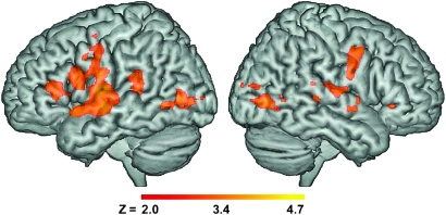
Understanding the neural mechanism that supports preserved language processing in aphasia has implications for both basic and applied science. This study examined brain activation associated with correct picture naming in 15 patients with aphasia. We contrasted each patient's activation to the activation observed in a neurologically healthy control group, allowing us to identify regions with unusual activity patterns. The results revealed that increased activation in preserved left hemisphere areas is associated with better naming performance in aphasia. This relationship was linear in nature; progressively less cortical activation was associated with greater severity of anomia. These findings are consistent with others who suggests that residual language function following stroke relies on preserved cortical areas in the left hemisphere.
Figures


References
-
- Basso A, Gardelli M, Grassi MP, Mariotti M. The role of the right-hemisphere in recovery from aphasia—2 case studies. Cortex. 1989;25:555–566. - PubMed
-
- Bates E, Wilson SM, Saygin AP, Dick F, Sereno MI, Knight RT, Dronkers NF. Voxel-based lesion-symptom mapping. Nat Neurosci. 2003;6:448–450. - PubMed
-
- Botvinick M, Nystrom LE, Fissell K, Carter CS, Cohen JD. Conflict monitoring versus selection-for-action in anterior cingulate cortex. Nature. 1999;402:179–181. - PubMed
-
- Breier JI, Castillo EM, Boake C, Billingsley R, Maher L, Francisco G, Papanicolaou AC. Spatiotemporal patterns of language-specific brain activity in patients with chronic aphasia after stroke using magnetoencephalography. Neuroimage. 2004;23:1308–1316. - PubMed
-
- Brett M, Leff AP, Rorden C, Ashburner J. Spatial normalization of brain images with focal lesions using cost function masking. Neuroimage. 2001;14:486–500. - PubMed
Publication types
MeSH terms
Substances
Grants and funding
LinkOut - more resources
Full Text Sources
Medical
