. 2009 Oct;29(20):5413-25.
doi: 10.1128/MCB.00368-09. Epub 2009 Aug 17.

FoxA1 binding directs chromatin structure and the functional response of a glucocorticoid receptor-regulated promoter

Affiliations

FoxA1 binding directs chromatin structure and the functional response of a glucocorticoid receptor-regulated promoter

Sergey Belikov et al. Mol Cell Biol. 2009 Oct.

Abstract

Reconstitution of the glucocorticoid receptor (GR)-regulated mouse mammary tumor virus (MMTV) promoter in Xenopus oocytes was used to monitor the effects of different transcription factor contexts. Three constitutively binding factors, nuclear factor 1 (NF1), octamer transcription factor 1 (Oct1), and the Forkhead box A1 (FoxA1), were shown to act in concert, to direct the chromatin structure, and to enhance the GR response. FoxA1 has a dominant effect in the absence of hormone and induces a cluster of DNase I-hypersensitive sites in the segment comprising bp -400 to +25. This FoxA1-mediated chromatin remodeling does not induce MMTV transcription, as opposed to that of the GR. However, the robust FoxA1-dependent chromatin opening has the following drastic functional consequences on the hormone regulation: (i) GR-DNA binding is facilitated, as revealed by dimethyl sulfate in vivo footprinting, leading to increased hormone-induced transcription, and (ii) the GR antagonist RU486 is converted into a partial agonist in the presence of FoxA1 via ligand-independent GR activation. We conclude that FoxA1 mediates a preset chromatin structure and directs a context-specific response of a nuclear receptor. Furthermore, the alternative nucleosome arrangement induced by GR and FoxA1 implies this to be determined by constitutive binding of transcription factors rather than by the DNA sequence itself.


Figures
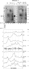
FIG. 1.
FIG. 1.
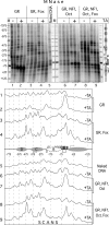
FoxA1-dependent chromatin remodeling of the MMTV LTR by DNase I and indirect end labeling. Oocytes injected with mRNA mixes coding for the indicated proteins and with single-stranded MMTV reporter DNA and treated or not treated with hormone (TA). Lane M, internal molecular weight markers. A summary of putative nucleosome positions and PhosphorImager scans of corresponding lanes are shown, and putative nucleosome positions in the hormone-activated state (5) and DNA binding sites are indicated, with the latter shown as boxes, for GR (white above the line), NF1 (gray), Oct1 (black), and FoxA1 (white below the line).
FIG. 2.
FIG. 2.
The distal FoxA1-dependent DHS harbors two FoxA1 binding sites. (A) Isolated DNAs from the experiment described in the legend to Fig. 1. DNase I digestion developed with primer extension using the −97/−123 primer. Positions of FoxA1 binding sequences, white boxes on the left. Scans of corresponding lanes are on the right. The characteristic hypersensitivity to DNase I induced by FoxA1 binding is highlighted (black arrowheads). (B) DMS methylation protection analysis and FoxA1-dependent methylation protection at the −333 and −224 sites, demonstrating binding. Oocytes were injected as described in the legend to Fig. 1 and assayed in duplicate (10 oocytes assayed two times). The digestion pattern was developed by a 33P-labeled primer extension, the −97/−123 primer. Scans of the corresponding lanes are shown below. Black arrowheads highlight the protected bands. (C) Schematic map and sequence ranging from −386 to −323 of the MMTV LTR. FoxA1 binding sites are underlined. Black arrowheads denote DHSs on the upper DNA strand. Bottom, FoxA1 consensus binding sequence adjusted from reference and sites confirmed by DNase I hypersensitivity and DMS methylation protection in the MMTV LTR.
FIG. 3.
FIG. 3.
Band shift with a DNA probe harboring the double FoxA1 sites at −360 and −332 demonstrates two FoxA1 mRNA injection (inj.)-dependent complexes (I and II with arrows on the left) that are both supershifted by a FoxA1 antibody (Ab) (marked with empty circles in lanes 5 and 12). Oocytes were either not injected (−) or injected (+) with FoxA1 mRNA 24 h prior to nuclear isolation. Oocyte nuclear extracts were prebound to a single-stranded oligonucleotide (Comp.), 1.7 μg (+) or 4.6 μg (+++) per sample, to compete for nonspecific DNA binding. α, anti-.
FIG. 4.
FIG. 4.
Nucleosome organization along the MMTV LTR is drastically affected by FoxA1, as revealed by MNase and indirect end labeling. Oocytes were injected as described in the legend to Fig. 1. Positions of protein binding sites are given on the right, as shown in Fig. 1. The scans and putative hormone-induced positions of nucleosomes in the MMTV LTR are shown below. Lane M, external molecular weight markers.
FIG. 5.
FIG. 5.
Effect of different transcription factor contexts on nucleosome organization analyzed by MNase and developed by the +92/+65 primer extension. The digestion pattern of isolated DNAs is from the experiment described in the legend to Fig. 3. Protein binding sites and the schematic representation of nucleosomal organization in oocytes containing GR alone or GR, NF1, and Oct1 are shown on the left. The schematic representation of nucleosomal organization of the MMTV promoter in oocytes containing GR and FoxA1 or GR, FoxA1, NF1, and Oct1 is shown on the right. The positions of the DNA segments protected by NF1 are highlighted by white arrowheads.
FIG. 6.
FIG. 6.
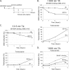
FoxA1 facilitates GR binding to DNA and enhances hormone-induced transcription, especially at low concentrations of hormone-activated GR. (A) Experimental design. (B) DMS in vivo footprinting analysis of GR-GRE binding and MMTV transcription analysis by S1 nuclease protection of oocytes treated with a high hormone (TA) concentration, i.e., 1,000 nM, plotted as a function of time. Double samples, 10 oocytes assayed two times, were analyzed for each time point. (C) Same as described in the legend to panel B, but a low hormone concentration was used, 10.5 nM TA. (D) Coexpression of NF1 and Oct1 together with GR and FoxA1 enhances hormone-induced transcription of the MMTV LTR. Individual values are indicated as black dots in the diagrams.
FIG. 7.
FIG. 7.
FoxA1 alters the RU486 antagonistic response into a partial agonist response. Oocytes, 2 × 10 in each group, were injected (inj) with mRNA to express proteins as indicated, followed by a single-stranded MMTV DNA reporter. Oocytes were divided into six groups and treated with the indicated hormone agonist/antagonist for 6 h, followed by analysis for transcriptional activity by S1 nuclease protection (A and B) and for specific protein-DNA binding by DMS methylation protection (C to E). GR binding, white arrowheads; FoxA1 binding, gray arrowhead; reference for loading control, black arrows. Bars shown in panels B, D, and E represent the average results from two independent analyses (black dots). Cort., corticosterone.
FIG. 8.
FIG. 8.
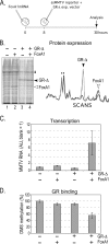
A truncated GR-Δ, devoid of the LBD, requires FoxA1 to bind its cognate DNA site and to induce MMTV transcription. (A, B) Oocytes for each group were injected as indicated (A) and analyzed for protein expression by SDS-PAGE (B), where PhosphorImager scans of lane 2 (black), lane 3 (dark gray), and lane 4 (light gray) indicate similar levels of the expressed proteins among the groups (right). (C, D) Transcription was analyzed by S1 nuclease protection in eight oocytes three times (C), and DNA binding was analyzed by DMS in vivo footprinting in eight oocytes three times (D) (as described in the legend to Fig. 7C). Standard deviations are indicated as error bars based on the results of triplicate samples.

References

    1. Astrand, C., S. Belikov, and O. Wrange. 2009. Histone acetylation characterizes chromatin presetting by NF1 and Oct1 and enhances glucocorticoid receptor binding to the MMTV promoter. Exp. Cell Res. 315:2604-2615. - PubMed
    1. Badis, G., E. T. Chan, H. van Bakel, L. Pena-Castillo, D. Tillo, K. Tsui, C. D. Carlson, A. J. Gossett, M. J. Hasinoff, C. L. Warren, M. Gebbia, S. Talukder, A. Yang, S. Mnaimneh, D. Terterov, D. Coburn, A. Li Yeo, Z. X. Yeo, N. D. Clarke, J. D. Lieb, A. Z. Ansari, C. Nislow, and T. R. Hughes. 2008. A library of yeast transcription factor motifs reveals a widespread function for Rsc3 in targeting nucleosome exclusion at promoters. Mol. Cell 32:878-887. - PMC - PubMed
    1. Belikov, S., C. Astrand, P. H. Holmqvist, and O. Wrange. 2004. Chromatin-mediated restriction of nuclear factor 1/CTF binding in a repressed and hormone-activated promoter in vivo. Mol. Cell. Biol. 24:3036-3047. - PMC - PubMed
    1. Belikov, S., C. Astrand, and O. Wrange. 2007. Mechanism of histone H1-stimulated glucocorticoid receptor DNA binding in vivo. Mol. Cell. Biol. 27:2398-2410. - PMC - PubMed
    1. Belikov, S., B. Gelius, G. Almouzni, and Ö. Wrange. 2000. Hormone activation induces nucleosome positioning in vivo. EMBO J. 19:1023-1033. - PMC - PubMed

Publication types