

Isolation and characterization of canine umbilical cord blood-derived mesenchymal stem cells
- PMID: 19687617
- PMCID: PMC2801133
- DOI: 10.4142/jvs.2009.10.3.181
Isolation and characterization of canine umbilical cord blood-derived mesenchymal stem cells
Erratum in
- J Vet Sci. 2009 Dec;10(4):369
Abstract
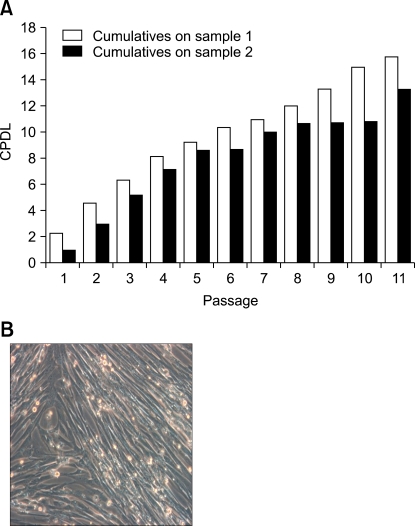
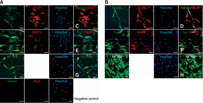
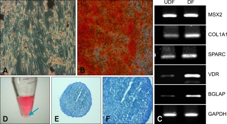
Human umbilical cord blood-derived mesenchymal stem cells (MSCs) are known to possess the potential for multiple differentiations abilities in vitro and in vivo. In canine system, studying stem cell therapy is important, but so far, stem cells from canine were not identified and characterized. In this study, we successfully isolated and characterized MSCs from the canine umbilical cord and its fetal blood. Canine MSCs (cMSCs) were grown in medium containing low glucose DMEM with 20% FBS. The cMSCs have stem cells expression patterns which are concerned with MSCs surface markers by fluorescence- activated cell sorter analysis. The cMSCs had multipotent abilities. In the neuronal differentiation study, the cMSCs expressed the neuronal markers glial fibrillary acidic protein (GFAP), neuronal class III beta tubulin (Tuj-1), neurofilament M (NF160) in the basal culture media. After neuronal differentiation, the cMSCs expressed the neuronal markers Nestin, GFAP, Tuj-1, microtubule-associated protein 2, NF160. In the osteogenic & chondrogenic differentiation studies, cMSCs were stained with alizarin red and toluidine blue staining, respectively. With osteogenic differentiation, the cMSCs presented osteoblastic differentiation genes by RT-PCR. This finding also suggests that cMSCs might have the ability to differentiate multipotentially. It was concluded that isolated MSCs from canine cord blood have multipotential differentiation abilities. Therefore, it is suggested that cMSCs may represent a be a good model system for stem cell biology and could be useful as a therapeutic modality for canine incurable or intractable diseases, including spinal cord injuries in future regenerative medicine studies.
Figures
Similar articles
-
Optimizing In Vitro Osteogenesis in Canine Autologous and Induced Pluripotent Stem Cell-Derived Mesenchymal Stromal Cells with Dexamethasone and BMP-2.Stem Cells Dev. 2021 Feb;30(4):214-226. doi: 10.1089/scd.2020.0144. Epub 2021 Feb 8. Stem Cells Dev. 2021. PMID: 33356875 Free PMC article.
-
The bone regenerative capacity of canine mesenchymal stem cells is regulated by site-specific multilineage differentiation.Oral Surg Oral Med Oral Pathol Oral Radiol. 2017 Feb;123(2):163-172. doi: 10.1016/j.oooo.2016.09.011. Epub 2016 Sep 28. Oral Surg Oral Med Oral Pathol Oral Radiol. 2017. PMID: 27876576 Free PMC article.
-
Chondrogenesis, osteogenesis and adipogenesis of canine mesenchymal stem cells: a biochemical, morphological and ultrastructural study.Histochem Cell Biol. 2007 Dec;128(6):507-20. doi: 10.1007/s00418-007-0337-z. Epub 2007 Oct 6. Histochem Cell Biol. 2007. PMID: 17922135
-
In-vitro characterization of canine multipotent stromal cells isolated from synovium, bone marrow, and adipose tissue: a donor-matched comparative study.Stem Cell Res Ther. 2017 Oct 3;8(1):218. doi: 10.1186/s13287-017-0639-6. Stem Cell Res Ther. 2017. PMID: 28974260 Free PMC article.
-
Donor-matched functional and molecular characterization of canine mesenchymal stem cells derived from different origins.Cell Transplant. 2013;22(12):2311-21. doi: 10.3727/096368912X657981. Epub 2012 Oct 12. Cell Transplant. 2013. PMID: 23068964
Cited by
-
Isolation and characterization of canine amniotic membrane-derived multipotent stem cells.PLoS One. 2012;7(9):e44693. doi: 10.1371/journal.pone.0044693. Epub 2012 Sep 14. PLoS One. 2012. PMID: 23024756 Free PMC article.
-
Canine Platelet Lysate Is Inferior to Fetal Bovine Serum for the Isolation and Propagation of Canine Adipose Tissue- and Bone Marrow-Derived Mesenchymal Stromal Cells.PLoS One. 2015 Sep 9;10(9):e0136621. doi: 10.1371/journal.pone.0136621. eCollection 2015. PLoS One. 2015. PMID: 26353112 Free PMC article.
-
A Comparative Study of Growth Kinetics, In Vitro Differentiation Potential and Molecular Characterization of Fetal Adnexa Derived Caprine Mesenchymal Stem Cells.PLoS One. 2016 Jun 3;11(6):e0156821. doi: 10.1371/journal.pone.0156821. eCollection 2016. PLoS One. 2016. PMID: 27257959 Free PMC article.
-
Durable Control of Autoimmune Diabetes in Mice Achieved by Intraperitoneal Transplantation of "Neo-Islets," Three-Dimensional Aggregates of Allogeneic Islet and "Mesenchymal Stem Cells".Stem Cells Transl Med. 2017 Jul;6(7):1631-1643. doi: 10.1002/sctm.17-0005. Epub 2017 May 3. Stem Cells Transl Med. 2017. PMID: 28467694 Free PMC article.
-
Stem cell therapy targets the neointimal smooth muscle cells in experimentally induced atherosclerosis: involvement of intracellular adhesion molecule (ICAM) and vascular cell adhesion molecule (VCAM).Braz J Med Biol Res. 2021 May 24;54(8):e10807. doi: 10.1590/1414-431X2020e10807. eCollection 2021. Braz J Med Biol Res. 2021. PMID: 34037094 Free PMC article.
References
-
- Bartholomew A, Sturgeon C, Siatskas M, Ferrer K, McIntosh K, Patil S, Hardy W, Devine S, Ucker D, Deans R, Moseley A, Hoffman R. Mesenchymal stem cells suppress lymphocyte proliferation in vitro and prolong skin graft survival in vivo. Exp Hematol. 2002;30:42–48. - PubMed
-
- Bernard BA. Human skin stem cells. J Soc Biol. 2008;202:3–6. - PubMed
-
- Bhattacharya V, McSweeney PA, Shi Q, Bruno B, Ishida A, Nash R, Storb RF, Sauvage LR, Hammond WP, Wu MH. Enhanced endothelialization and microvessel formation in polyester grafts seeded with CD34(+) bone marrow cells. Blood. 2000;95:581–585. - PubMed
-
- Bieback K, Kern S, Klüter H, Eichler H. Critical parameters for the isolation of mesenchymal stem cells from umbilical cord blood. Stem Cells. 2004;22:625–634. - PubMed
-
- Breems DA, Löwenberg B. Acute myeloid leukemia and the position of autologous stem cell transplantation. Semin Hematol. 2007;44:259–266. - PubMed
Publication types
MeSH terms
LinkOut - more resources
Full Text Sources
Other Literature Sources
Miscellaneous
