Role of protease inhibitors and acylation stimulating protein in the adipogenesis in 3T3-L1 cells
- PMID: 19687619
- PMCID: PMC2801122
- DOI: 10.4142/jvs.2009.10.3.197
Role of protease inhibitors and acylation stimulating protein in the adipogenesis in 3T3-L1 cells
Abstract
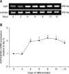
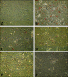
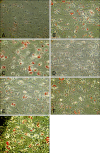
Treatment of AIDS (HIV) and hepatitis C virus needs protease inhibitors (PI) to prevent viral replication. Uses of PI in therapy are usually associated with a decrease in body weight and dyslipidemia. Acylation stimulating protein (ASP) is a protein synthesized in adipocytes to increase triglycerides biosynthesis, for that the relation of PI and ASP to adipogenesis is tested in this work. ASP expression was increased during 3T3-L1 differentiation and reached a peak at day 8 with cell maturation. Addition of PI during adipocytes differentiation dose dependently and significantly (p < 0.5) inhibited the degree of triglycerides (TG) accumulation. Moreover, presence of ASP (450 ng/mL) in media significantly (p < 0.5) stimulated the degree of TG accumulation and there was additive stimulation for ASP when added with insulin (10 microg/mL). Finally, when ASP in different doses (Low, 16.7; Medium, 45 and High, 450 ng/mL) incubated with a dose of x150 PI, ASP partially inhibited the PI-inhibited adipogenesis and TG accumulation. The results in this study show that PI inhibit lipids accumulation and confirm role of ASP in TG biosynthesis and adipogenesis.
Figures


Similar articles
-
Evaluation of chylomicron effect on ASP production in 3T3-L1 adipocytes.Acta Biochim Biophys Sin (Shanghai). 2011 Feb;43(2):154-9. doi: 10.1093/abbs/gmq124. Acta Biochim Biophys Sin (Shanghai). 2011. PMID: 21266544
-
[Effect of metabolic drugs on the secretion of acylation stimulating protein in 3T3-L1 adipocytes].Xi Bao Yu Fen Zi Mian Yi Xue Za Zhi. 2012 Oct;28(10):1051-4. Xi Bao Yu Fen Zi Mian Yi Xue Za Zhi. 2012. PMID: 23046937 Chinese.
-
Differential chemoattractant response in adipocytes and macrophages to the action of acylation stimulating protein.Eur J Cell Biol. 2013 Feb;92(2):61-9. doi: 10.1016/j.ejcb.2012.10.005. Epub 2012 Dec 14. Eur J Cell Biol. 2013. PMID: 23245988
-
[Effect of acylation stimulating protein on the perilipin and adipophilin expression during 3T3-L1 preadipocyte differentiation.].Sheng Li Xue Bao. 2009 Feb 25;61(1):56-64. Sheng Li Xue Bao. 2009. PMID: 19224055 Chinese.
-
Gly-Ala-Gly-Val-Gly-Tyr, a novel synthetic peptide, improves glucose transport and exerts beneficial lipid metabolic effects in 3T3-L1 adipoctyes.Eur J Pharmacol. 2011 Jan 10;650(1):479-85. doi: 10.1016/j.ejphar.2010.10.006. Epub 2010 Oct 14. Eur J Pharmacol. 2011. PMID: 20951125
Cited by
-
The function of adipsin and C9 protein in the complement system in HIV-associated preeclampsia.Arch Gynecol Obstet. 2021 Dec;304(6):1467-1473. doi: 10.1007/s00404-021-06069-9. Epub 2021 Apr 21. Arch Gynecol Obstet. 2021. PMID: 33881585
References
-
- Berger J, Leibowitz MD, Doebber TW, Elbrecht A, Zhang B, Zhou G, Biswas C, Cullinan CA, Hayes NS, Li Y, Tanen M, Ventre J, Wu MS, Berger GD, Mosley R, Marquis R, Santini C, Sahoo SP, Tolman RL, Smith RG, Moller DE. Novel peroxisome proliferator-activated receptor (PPAR) γ and PPARδ ligands produce distinct biological effects. J Bio Chem. 1999;274:6718–6725. - PubMed
-
- Caron M, Auclair M, Vigouroux C, Glorian M, Forest C, Capeau J. The HIV protease inhibitor indinavir impairs sterol regulatory element-binding protein-1 intranuclear localization, inhibits preadipocyte differentiation, and induces insulin resistance. Diabetes. 2001;50:1378–1388. - PubMed
-
- Carr A, Samaras K, Burton S, Law M, Freund J, Chisholm DJ, Cooper DA. A syndrome of peripheral lipodystrophy, hyperlipidaemia and insulin resistance in patients receiving HIV protease inhibitors. AIDS. 1998;12:F51–F58. - PubMed
-
- Carr A, Samaras K, Chisholm DJ, Cooper DA. Pathogenesis of HIV-1-protease inhibitor-associated peripheral lipodystrophy, hyperlipidaemia, and insulin resistance. Lancet. 1998;351:1881–1883. - PubMed
-
- Choy LN, Rosen BS, Spiegelman BM. Adipsin and an endogenous pathway of complement from adipose cells. J Biol Chem. 1992;267:12736–12741. - PubMed
MeSH terms
Substances
LinkOut - more resources
Full Text Sources
Research Materials
Miscellaneous

