

Chromophore concentrations, absorption and scattering properties of human skin in-vivo
- PMID: 19687939
- PMCID: PMC2754563
- DOI: 10.1364/oe.17.014599
Chromophore concentrations, absorption and scattering properties of human skin in-vivo
Abstract
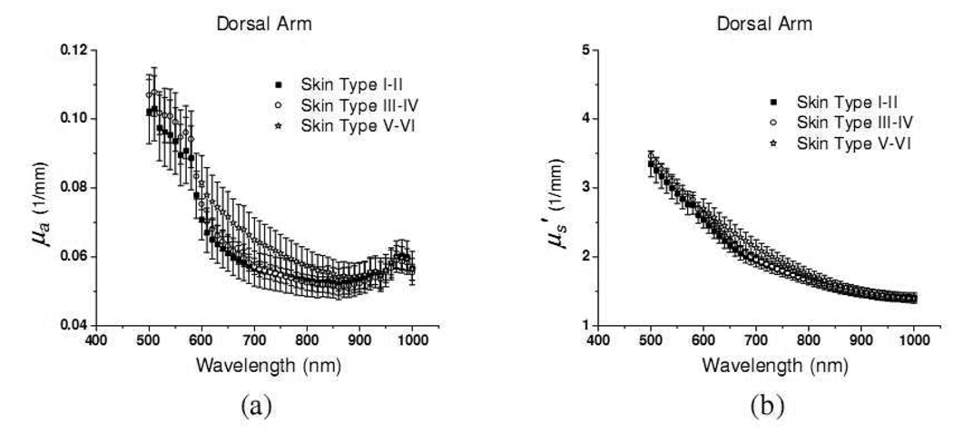
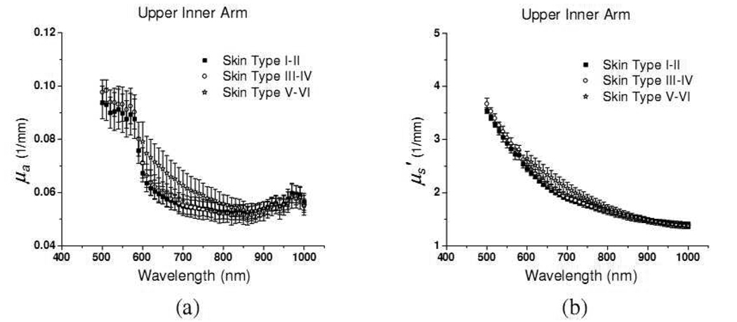
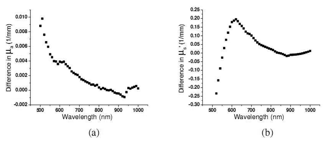
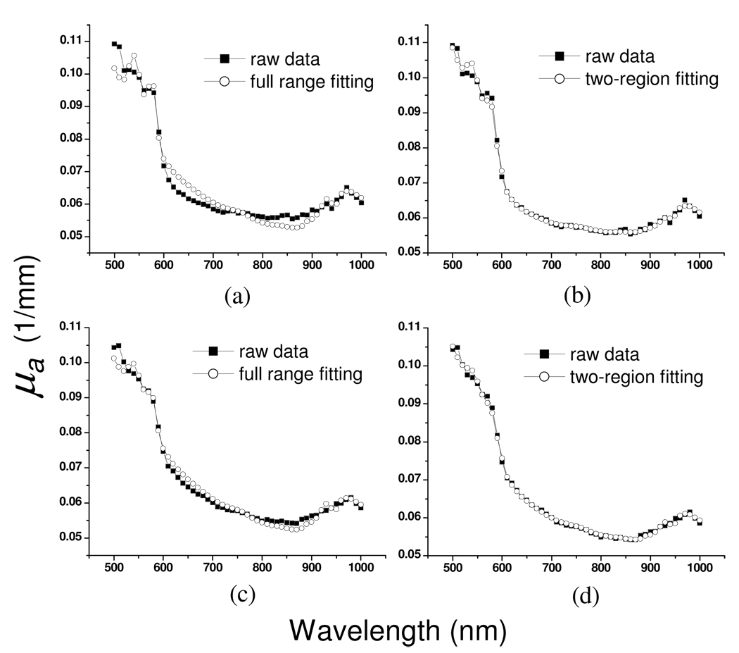
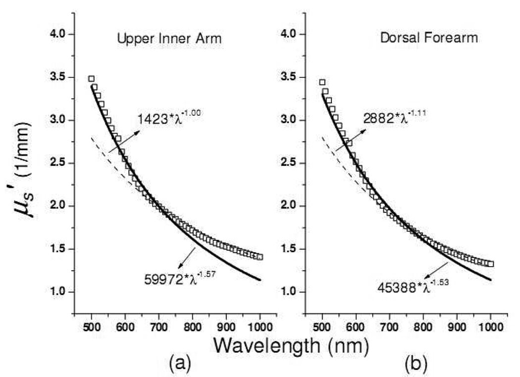
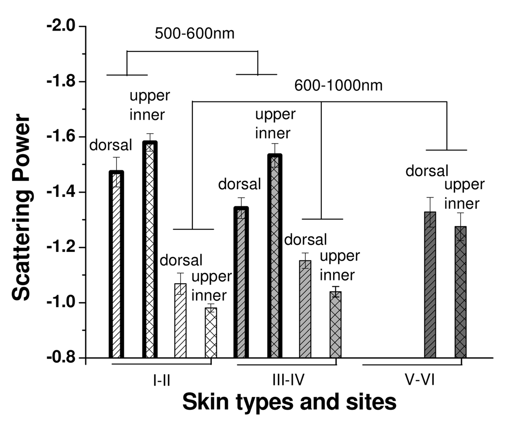
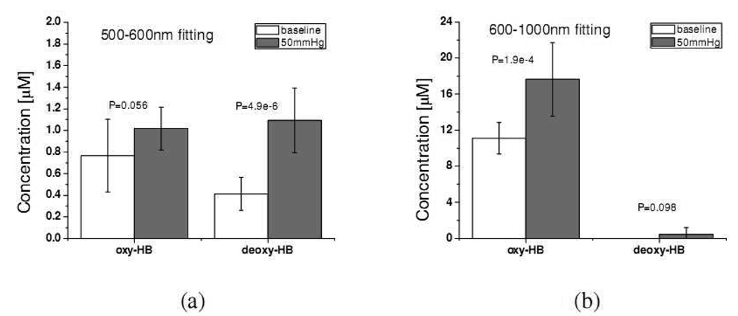
Absorption and reduced scattering coefficients of in-vivo human skin provide critical information on non-invasive skin diagnoses for aesthetic and clinical purposes. To date, very few in-vivo skin optical properties have been reported. Previously, we reported absorption and scattering properties of in-vivo skin in the wavelength range from 650 to 1000 nm using the diffusing probe in the "modified two-layer geometry". In this study, we determine the spectra of skin optical properties continuously in the range from 500 to 1000 nm. It was found that the concentration of chromophores, such as oxy-hemoglobin, deoxy-hemoglobin, and melanin, calculated based on the absorption spectra of eighteen subjects at wavelengths above and below 600 nm were distinct because of the inherent difference in the interrogation region. The scattering power, which is related to the average scatterer's size, demonstrates a clear contrast between skin phototypes, skin sites, and wavelengths. We also applied venous occlusion on forearms and found that the concentrations of oxy- and deoxy-hemoglobin as assessed at wavelengths above and below 600 nm were different. Our results suggest that diffuse reflectance techniques with the visible and near infrared light sources can be employed to investigate the hemodynamics and optical properties of upper dermis and lower dermis.
Figures
References
-
- Nishidate I, Aizu Y, Mishina H. Estimation of melanin and hemoglobin in skin tissue using multiple regression analysis aided by Monte Carlo simulation. J. Biomed. Opt. 2004;9:700–710. - PubMed
-
- Chang SK, Arifler D, Drezek R, Follen M, Richards-Kortum R. Analytical model to describe fluorescence spectra of normal and preneoplastic epithelial tissue: comparison with Monte Carlo simulations and clinical measurements. J. Biomed. Opt. 2004;9:511–522. - PubMed
-
- Kelly KM, Choi B, McFarlane S, Motosue A, Jung BJ, Khan MH, Ramirez-San-Juan JC, Nelson JS. Description and analysis of treatments for port-wine stain birthmarks. Arch. Facial Plast. Surg. 2005;7:287–294. - PubMed
-
- Tromberg BJ, Svaasand LO, Fehr MK, Madsen SJ, Wyss P, Sansone B, Tadir Y. A mathematical model for light dosimetry in photodynamic destruction of human endometrium. Phys. Med. Biol. 1996;41:223–237. - PubMed
-
- Troy TL, Thennadil SN. Optical properties of human skin in the near infrared wavelength range of 1000 to 2200 nm. J. Biomed. Opt. 2001;6:167–176. - PubMed
Publication types
MeSH terms
Substances
Grants and funding
LinkOut - more resources
Full Text Sources
Other Literature Sources
