Review
. 2010 Jan;40(1-2):143-53.
doi: 10.1007/s12031-009-9229-0. Epub 2009 Aug 18.

What have we learned from the congenital myasthenic syndromes

Affiliations
Review

What have we learned from the congenital myasthenic syndromes

Andrew G Engel et al. J Mol Neurosci. 2010 Jan.

Abstract

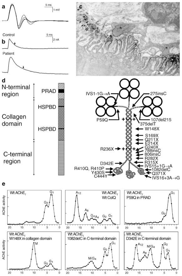
The congenital myasthenic syndromes have now been traced to an array of molecular targets at the neuromuscular junction encoded by no fewer than 11 disease genes. The disease genes were identified by the candidate gene approach, using clues derived from clinical, electrophysiological, cytochemical, and ultrastructural features. For example, electrophysiologic studies in patients suffering from sudden episodes of apnea pointed to a defect in acetylcholine resynthesis and CHAT as the candidate gene (Ohno et al., Proc Natl Acad Sci USA 98:2017-2022, 2001); refractoriness to anticholinesterase medications and partial or complete absence of acetylcholinesterase (AChE) from the endplates (EPs) has pointed to one of the two genes (COLQ and ACHE ( T )) encoding AChE, though mutations were observed only in COLQ. After a series of patients carrying mutations in a disease gene have been identified, the emerging genotype-phenotype correlations provided clues for targeted mutation analysis in other patients. Mutations in EP-specific proteins also prompted expression studies that proved pathogenicity, highlighted important functional domains of the abnormal proteins, and pointed to rational therapy.


Figures

Figure 1
Figure 1
CMS caused by defects in AChE. a Repetitive CMAPs are generated by prolonged synaptic potentials that outlast the absolute refractory period of the muscle fiber. b MEPCs recorded from a normal and an AChE-deficient patient EP. Vertical arrows indicate decay time constants. The patient MEPC decays slowly as ACh repeatedly binds to AChRs before it exits the synaptic space by diffusion; the MEPC amplitude is reduced owing to the associated EP myopathy. c AChE-deficient EP region. AChR is localized with peroxidase-labeled α-bungarotoxin. Numerous junctional folds are degenerating, shedding their terminal expansions into the synaptic space. The nerve terminals are abnormally small and cover only a fraction of the postsynaptic region. d Schematic diagram showing domains of a ColQ strand and components of the A12 species of asymmetric AChE with 24 identified ColQ mutations. e Clockwise from upper left density gradient ultracentrifugation profiles of extracts of COS cells transfected with wild-type AChET, wild-type AChET and wild-type COLQ, wild-type AChET and the P59Q PRAD domain mutant of COLQ, wild-type AChET and the W148X collagen domain mutant of COLQ; wild-type AChET and the 1082delC C-terminal domain mutant of COLQ; and wild-type AChET and the D342E C-terminal domain mutant of COLQ. Note that the asymmetric forms of AChE appear only after transfection with the C-terminal D342E mutant of COLQ. AChE acetylcholinesterase, MEPC miniature EP current, HSPBD heparan sulfate proteoglycan binding domain, PRAD proline-rich attachment domain, Wt wild type, G globular moiety of AChET, A asymmetric moiety of AChE, M mutant peak. (Reproduced by permission from Engel et al. 2003b)
Figure 2
Figure 2
ChAT mutations in CMS patients mapped on stereo image of structural model of rat ChAT (PDB code1Q6X). The active site is in a solvent-accessible tunnel at the interface of the N (upper) and C (lower) domains. Histidine at the active site is shown in stick representation. Residue numbers correspond to the human sequence. The indicated mutations, except those in boxes, were identified in our laboratory
Figure 3
Figure 3
Slow-channel syndromes. a Schematic diagram of AChR subunits with slow-channel mutation. b Single-channel currents from wild-type and slow-channel (αV249F) AChRs expressed in HEK cells. c MEPC recorded from EPs of a control subject and a patient harboring the αV249F slow-channel mutation. The slow-channel MEPC decays biexponentially due to expression of both wild-type and mutant AChRs at the EP. d Slow-channel EP. The junctional folds have disintegrated and the synaptic space is filled with debris (black asterisk). The junctional sarcoplasm displays fragmented apoptotic nuclei (white asterisk) and myeloid structures. Bar=1 μm. (a to c are from Engel 2004, by permission)
Figure 4
Figure 4
Fast-channel syndromes. a MEPC recorded from EPs of a control subject and a patient harboring the αV285I fast-channel mutation. Arrows indicate decay time constants. b Single-channel currents from wild-type and fast-channel (αV285I) AChRs expressed in HEK cells. c Schematic diagram of fast-channel mutations in the AChR α, β, and δ subunits. (From Engel 2004, by permission)
Figure 5
Figure 5
Kinetic scheme for activation of wild-type, εN436del, and δL42P AChR. AChR activation involves reversible binding of two molecules of agonist (A) to the AChR in the resting, closed state (R), followed by reversible formation of the open state (R*). K1 and K2 are equilibrium dissociation constants of the first and second agonist binding steps; θ indicates the gating equilibrium constant. Values indicated in gray differ markedly from corresponding values for the wild type
Figure 6
Figure 6
Structural model of the AChR α and δ subunits and mutant cycle analyses. a An enlarged view of the coupled intersubunit residues αY127 and δN41 in the structural model of the Torpedo AChR (Protein Data Bank code 2BG9). b A mutant cycle for the mutations αY127T, δL42P, and εL40P. Single-channel currents correspond to each AChR elicited by 100 μM ACh. Changes in gating free energy along each limb of the cycle are shown, and the overall coupling free energy (ΔΔGint) in units of kilocalories per mole computed from −RT ln[(θwwθmm)/(θwmθmw)] where the θ(β/α) is the gating equilibrium constant for diliganded receptors for wild-type, single-mutant, or double-mutant AChRs. Horizontal bar indicates 20 ms for wild-type AChRs and 100 ms for the mutant AChRs. Vertical bar indicates 5 pA. (Reproduced by permission from Shen et al. 2008)
Figure 7
Figure 7
Dok-7 myasthenia. a Degenerating EP region displaying loss or amputation of junctional folds and accumulation of degenerate fold remnants in the widened synaptic space. b Unstained normal EP region reacted for AChR with peroxidase conjugated α-bungarotoxin. The junctional folds are intact and display a normal concentration and distribution of AChR on the junctional fold. Bars=1 μm
Figure 8
Figure 8
Dok-7 myasthenia; EPs with presynaptic and postsynaptic abnormalities. a A highly abnormal EP showing extensive degeneration of the junctional folds (asterisks) and widening of the synaptic space. b This EP region shows extensive accumulation of myeloid structures in the junctional sarcoplasm. Some junctional folds are degenerating (asterisk). The postsynaptic region is denuded of its nerve terminal. A nerve sprout (s) surrounded by Schwann cell appears above the junction. Bars=1 μm

References

    1. Barisic N, Muller JS, Paucic-Kirincic E, et al. Clinical variability of CMS-EA (congenital myasthenic syndrome with episodic apnea) due to identical CHAT mutations in two infants. European Journal of Paediatric Neurology. 2005;9:7–12. - PubMed
    1. Beeson D, Hantai D, Lochmuller H, Engel AG. Report of the 126th International Workshop: Congenital myasthenic syndromes. Neuromuscular Disorders. 2005;15:498–512. - PubMed
    1. Beeson D, Higuchi O, Palace J, et al. Dok-7 mutations underlie a neuromuscular junction synaptopathy. Science. 2006;313:1975–1978. - PubMed
    1. Bon S, Coussen F, Massoulié J. Quaternary associations of acetylcholinesterase. II. The polyproline attachment domain of the collagen tail. Journal of Biological Chemistry. 1997;272:3016–3021. - PubMed
    1. Byring RF, Pihko H, Shen XM, et al. Congenital myasthenic syndrome associated with episodic apnea and sudden infant death. Neuromuscular Disorders. 2002;12:548–553. - PubMed

Publication types

MeSH terms

LinkOut - more resources