Review
. 2009 Nov 20;4(11):895-909.
doi: 10.1021/cb900163d.

Flipping lipids: why an' what's the reason for?

Affiliations
Review

Flipping lipids: why an' what's the reason for?

Sumana Sanyal et al. ACS Chem Biol. .

Abstract

The biosynthesis of glycoconjugates such as N-glycoproteins and GPI-anchored proteins in eukaryotes and cell wall peptidoglycan and lipopolysaccharide in bacteria requires lipid intermediates to be flipped rapidly across the endoplasmic reticulum or bacterial cytoplasmic membrane (so-called biogenic membranes). Rapid flipping is also required to normalize the number of glycerophospholipids in the two leaflets of the bilayer as the membrane expands in a growing cell. Although lipids diffuse rapidly in the plane of the membrane, the intrinsic rate at which they flip across membranes is very low. Biogenic membranes possess dedicated lipid transporters or flippases to increase flipping to a physiologically sufficient rate. The flippases are "ATP-independent" and facilitate "downhill" transport. Most predicted biogenic membrane flippases have not been identified at the molecular level, and the few flippases that have been identified by genetic approaches have not been biochemically validated. Here we summarize recent progress on this fundamental topic and speculate on the mechanism(s) by which biogenic membrane flippases facilitate transbilayer lipid movement.


Figures

Figure 1
Figure 1. Glycerophospholipid synthesis and flip-flop at the ER
(A) Phosphatidylcholine (PC), a major glycerophospholipid in eukaryotic cells. (B) A glycerophospholipid (PL) biosynthetic enzyme (left) converts diacylglycerol to PL on the cytoplasmic face of the ER. The PL flippase (right) facilitates bidirectional transbilayer translocation of PLs, allowing balanced growth of the bilayer. The ER PL flippase does not itself generate or maintain transbilayer lipid asymmetry. (C) Shape change induced in a prolate giant unilamellar vesicle (left) by the introduction of 0.1 mol percent lyso PC into the outer leaflet. Scale bar, 10 µm. Image courtesy of A. Papadopulos.
Figure 2
Figure 2. Protein N-glycosylation and GPI anchoring in the ER
(A) Oligosaccharyltransferase (OST) transfers Glc3Man9GlcNAc2 from Glc3Man9GlcNAc2-PP-dolichol (left) to an asparagine residue (yellow) within a glycosylation sequon (Asn-X-Ser/Thr) in the nascent polypeptide (right) as it emerges into the ER lumen. (B) An ER-translocated protein with a C-terminal GPI-directing signal sequence (orange) is a substrate for GPI transamidase (GPIT). GPIT replaces the C-terminal signal sequence with a GPI anchor. The anchor is attached via an amide bond between an ethanolamine residue in the GPI and the α-carboxyl group of the C-terminal amino acid. Symbols are explained in Fig. 3. (C) Structure of Glc3Man9GlcNAc2-PP-dolichol (a 75-carbon yeast dolichol is depicted) and the mammalian GPI H8.
Figure 3
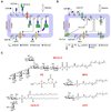
Figure 3. Lipid flipping in protein N-glycosylation and GPI anchoring in the ER
(A) Assembly of Glc3Man9GlcNAc2-PP-dolichol. The first 7 steps, leading to the synthesis of M5-DLO from dolichyl phosphate occur on the cytoplasmic face of the ER. GDP-mannose and UDP-GlcNAc directly contribute the sugar moieties for these reactions. M5-DLO is then flipped into the ER lumen by M5-DLO flippase, and extended in a further 7 reactions to yield Glc3Man9GlcNAc2-PP-dolichol. The sugar donors for these lumenal reactions are MPD and GPD. MPD is synthesized from dolichyl phosphate and GDP-mannose on the cytoplasmic face of the ER, then flipped to the ER lumen by MPD flippase. In the lumen it is the mannosyl donor for the 4 reactions that convert M5-DLO to Man9GlcNAc2-PP-dolichol. GPD is synthesized from dolichyl phosphate and UDP-glucose (not shown) on the cytoplasmic face of the ER, then flipped to the ER lumen by GPD flippase. (B) GPI biosynthesis. GlcN-PI is synthesized in two steps from PI on the cytoplasmic face of the ER, then flipped into the ER lumen to be elaborated into a mature GPI anchor. Lumenal reactions include inositol acylation, and mannose and phosphoethanolamine addition. The inositol acyl group is derived from fatty acyl CoA (FA-CoA) which is transported into the ER by an unknown mechanism. Mannose and phosphoethanolamine residues are derived from MPD and PE, respectively; both these lipids have to be flipped into the ER lumen. (C) Structures of the lipids that are flipped across the ER membrane in the dolichol cycle and GPI biosynthesis pathways. Yeast dolichol (15 isoprene units) is depicted in the M5-DLO, MPD and GPD structures; mammalian dolichol is longer, typically 19 isoprene units.
Figure 4
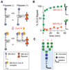
Figure 4. M5-DLO flipping in a reconstituted system
(A) Principle of the M5-DLO flippase assay in reconstituted vesicles. Vesicles are reconstituted with trace amounts of [3H]M5-DLO. Protein-free liposomes or vesicles containing irrelevant membrane proteins are indicated as Flippase (−); proteoliposomes containing the M5-DLO flippase are indicated Flippase (+). On adding the lectin Con A to the vesicles, M5-DLO molecules in the outer leaflet bind Con A and are rendered insoluble in an organic solvent (a mixture of chloroform, methanol and water); M5-DLO molecules in the inner leaflet are protected. Thus, for Flippase (−) vesicles, 50% of the M5-DLO molecules are captured by Con A; for Flippase (+) vesicles, 100% are captured since those in the inner leaflet are translocated to the outer leaflet. (B) Kinetics of flipping (obtained from the rate of capture of M5-DLO by Con A in intact vesicles (42)) of M5-DLO and its structural isomer, iM5-DLO. Symbols are explained in panel C. (C) The structure of Man9GlcNAc2-PP-dolichol is depicted. The M5-DLO sub-structure is contained within the light blue shaded region. M3-DLO (structure enclosed within the dashed lines) is flipped almost as well as M5-DLO. DLO structures containing mannoses indicated “+” are flipped more rapidly whereas the presence of mannose residues indicated by “−” reduces the rate of flipping (42). These rules indicate that M5-DLO is the optimal substrate for the DLO flippase.
Figure 5
Figure 5. Flipping of glycolipids across the bCM
(A) Peptidoglycan assembly. MurNAc-pentapeptide is transferred from UDP-MurNAc-pentapeptide to undecaprenyl phosphate to form Lipid I. Addition of GlcNAc from UDP-GlcNAc to Lipid I generates Lipid II on the cytoplasmic face of the bCM. Lipid II is translocated by a flippase to the exoplasmic/periplasmic surface. Here, its GlcNAc-MurNAc-peptide headgroup is transferred to a growing peptidoglycan polymer by transglycosylation, following which undecaprenyl diphosphate is recycled. Transpeptidation reactions link polymer chains to form the cell wall (orange bricks). Penicillin binding proteins (PBPs), many of which are bi-functional and contain both transglycosylase and transpeptidase activities, catalyze these later steps. (B) O-antigen and lipopolysaccharide assembly in Gram-negative bacteria. Hexa-acylated Lipid A, synthesized on the cytoplasmic surface of the bCM, is modified with 3-deoxy-d-manno-octulosonic Acid (Kdo) residues and a core oligosaccharide before being translocated to the periplasmic face by the ABC transporter MsbA. In parallel, the undecaprenyl diphosphate-linked repeat unit of the O-antigen is flipped across the bCM; the flippase is presumed to be a member of the Wzx family of proteins. The O-antigen units are polymerized as shown, then ligated to Lipid A-Kdo2-core. (C) Structure of Lipid II (shown with lysine in its pentapeptide side-chain; diaminopimelic acid is also commonly found instead of lysine), GlcNAc-PP-undecaprenol and aminoarabinose-P-undecaprenol and Lipid A-Kdo2-core oligosaccharide (decorated with O-antigen and aminoarabinose (in blue); these modifications to Lipid A-Kdo2-core oligosaccharide occur on the periplasmic face of the bCM). GlcNAc-PP-undecaprenol is recognized by the O-antigen assembly system, resulting in transfer of GlcNAc to lipid A-core (72). An analog of GlcNAc-PP-undecaprenol was used to test the role of WzxE as a flippase (71).
Figure 6
Figure 6. Flippase mechanisms
(A) A pore model for the PL flippase provides a central hydrophilic path for the transiting headgroup while leaving the hydrophobic chains of the lipid in the bilayer. The lipid is shown (in a number of snapshots) intercalating between transmembrane spans of the flippase. In this model the lipid is not specifically recognized, but conformationally constrained hydrophobic entities such as dolichol would prevent MPD or M5-DLO from intercalating into and being transported by the PL-flippase. (B) Slip-pop model for phospholipid transport. The dynamic behavior of hydrophobic single-membrane-spanning proteins causes transient defects in the lipid-helix interface that allow phospholipids to flip-flop across the bilayer. (C) Possible mechanism for the M5-DLO flippase (flippases for MPD (Fig. 3C) and other isoprenoid-based lipids such as Lipid II (Fig. 5C), may use a similar mechanism). Snapshots of a transiting M5-DLO molecule are shown. M5-DLO specifically binds to the flippase at one or other membrane interface, then exchanges with a symmetrically located binding site from which it is released into the bilayer. A single, centrally located binding site can also be envisaged, possibly located in a ‘thin’ portion of the membrane.

Similar articles

Cited by

References

    1. Palade GE. Membrane biogenesis: an overview. Methods Enzymol. 1983;96:xxix–lv. - PubMed
    1. Blobel G. Protein targeting (Nobel lecture) Chembiochem. 2000;1:86–102. - PubMed
    1. White SH, von Heijne G. How translocons select transmembrane helices. Annu Rev Biophys. 2008;37:23–42. - PubMed
    1. Kornberg RD, McConnell HM. Inside-outside transitions of phospholipids in vesicle membranes. Biochemistry. 1971;10:1111–1120. - PubMed
    1. Liu J, Conboy JC. 1,2-diacyl-phosphatidylcholine flip-flop measured directly by sum-frequency vibrational spectroscopy. Biophys J. 2005;89:2522–2532. - PMC - PubMed

Publication types