. 2009 Sep;128(1):25-33.
doi: 10.1111/j.1365-2567.2009.03046.x.

CX3CR1-deficiency is associated with increased severity of disease in experimental autoimmune uveitis

Affiliations

CX3CR1-deficiency is associated with increased severity of disease in experimental autoimmune uveitis

Athanasios Dagkalis et al. Immunology. 2009 Sep.

Abstract

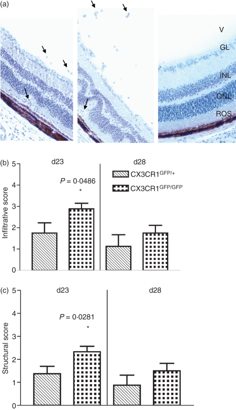
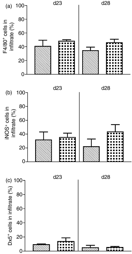
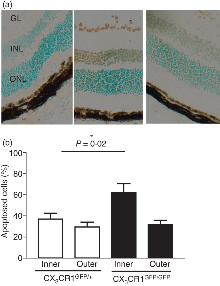
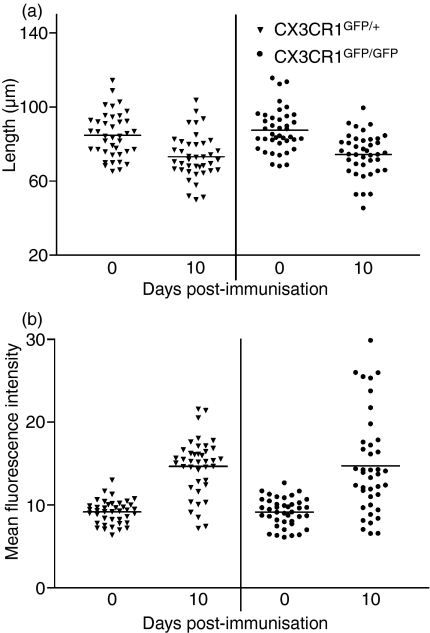
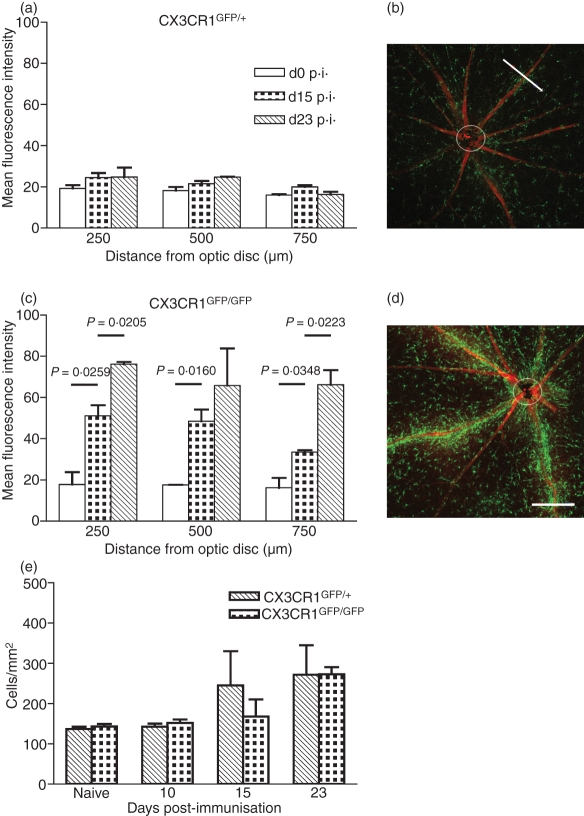
The role of CX3CR1 in regulating the function of monocytes and microglia was examined in mice in which CX3CR1 had been replaced by green fluorescent protein (GFP). Induction of experimental autoimmune uveitis (EAU) in these mice resulted in increased disease severity at day 23 postimmunization with uveitogenic peptide when compared with CX3CR1-positive mice and increased apoptosis of neuronal cells in the inner nuclear layer. Resident microglia within the retina were activated equally as EAU developed in mice with or without CX3CR1, as determined by changes in morphology, suggesting that the microglial cell response did not account for the differences. Although the inflammatory infiltrate had increased in mice without CX3CR1 at day 23 postimmunization, the percentage of natural killer cells in the infiltrate was not changed in these mice. Similarly, increased disease severity at this stage was not associated with an overall increased percentage of macrophages in the retinal inflammatory infiltrate or in increased activation of these cells. The increased recruitment of monocytes to the retina in response to EAU induction in CX3CR1(GFP/GFP) mice compared with CX3CR1(GFP/+) mice was not reflected in increased migration away from vessels, leading to marked clustering of GFP(+) cells around veins and venules in these mice. It is possible that this monocyte/macrophage clustering leads to the increased severity of disease seen in the mice by focusing and so intensifying the inflammatory response.


Figures

Figure 1
Figure 1
Effect of lack of CX3CR1 on experimental autoimmune uveitis (EAU). (a) Haematoxylin-stained cryostat sections from a CX3CR1GFP/+ mouse (left) and a CX3CR1GFP/GFP mouse (middle) both day 23 postimmunization (p.i.) and a CX3CR1GFP/GFP mouse non-immunized (right) (V, vitreous; GL, ganglion layer; INL, inner nuclear layer; ONL, outer nuclear layer; ROS, rod outer segments; arrows indicate examples of infiltration of inflammatory cells; original magnification × 25). (b) Disease graded by inflammatory infiltrate and (c) structural damage score in CX3CR1GFP/+ and CX3CR1GFP/GFP mice at days 23 and 28 p.i. Hatched bars, CX3CR1GFP/+ mice. Dotted bars, CX3CR1GFP/GFP mice. Error bars indicate SEM; n = 8 mice per group.
Figure 2
Figure 2
Effect of lack of CX3CR1 on cellular composition of inflammatory infiltrate in experimental autoimmune uveitis (EAU). (a) Percentage of cells in the inflammatory infiltrate stained positively for F4/80. (b) Percentage of cells in the inflammatory infiltrate stained positively for inducible nitric oxide synthase (iNOS). (c) Percentage of cells in the inflammatory infiltrate stained positively for DX5. Hatched bars, CX3CR1GFP/+ mice. Dotted bars, CX3CR1GFP/GFP mice. Error bars indicate SEM; n = 8 mice per group.
Figure 3
Figure 3
Effect of lack of CX3CR1 on apoptosis in nuclear layers of the retina in experimental autoimmune uveitis (EAU). (a) Cryostat sections from CX3CR1GFP/GFP, assay control (left section), from CX3CR1GFP/GFP TUNEL stained (middle section), and from CX3CR1GFP/+ TUNEL stained (right section), all day 23 postimmunization (p.i.) with apoptosis (brown stain) detected by TUNEL staining using a Klenow FragEL™ DNA fragmentation detection kit (GL, ganglion layer; INL, inner nuclear layer; ONL, outer nuclear layer; original magnification × 50). (b) Percentage of apoptosing cells in the inner and outer nuclear layer of the retina in CX3CR1GFP/+ (open bars) and CX3CR1GFP/GFP mice (filled bars) day 23 p.i. Error bars indicate SEM; n= 8 mice per group.
Figure 4
Figure 4
Activation of microglia during early experimental autoimmune uveitis (EAU) development to day 10 postimmunization (p.i.) in CX3CR1GFP/+ and CX3CR1GFP/GFP mice. (a) Average length of microglial dendrites in retinal whole-mounts. All visible dendrites (three or four) were measured per cell. (b) Mean fluorescence intensity (MFI) of individual microglia in retinal whole-mounts. Triangles, CX3CR1GFP/+ mice; circles, CX3CR1GFP/GFP mice. Error bars indicate SEM; n= 40 cells per group.
Figure 5
Figure 5
Effect of lack of CX3CR1 on distribution of green fluorescent protein (GFP)-positive cells around inflamed veins and venules. Mean fluorescence intensity (MFI) measured perpendicular to the length of the vessel at 250, 500 and 750 μm from the optic disc in retinal whole-mounts from (a) CX3CR1GFP/+ mice and (c) CX3CR1GFP/GFP mice. Open bars, no induction of experimental autoimmune uveitis (EAU); dotted bars, day 15 postimmunization (p.i.); hatched bars, day 23 p.i. Example of confocal image for CX3CR1GFP/+ and CX3CR1GFP/GFP mice day 23 p.i., (b) and (d), respectively, with white bar 180 μm indicating a measurement. Individual GFP-expressing cells counted between 100 and 500 μm away from the centre of the vessel at days 0, 10, 15 and day 23 p.i. (e) Hatched bars, CX3CR1GFP/+ mice. Dotted bars, CX3CR1GFP/GFP mice; n= 3 mice per group. Error bars indicate SEM.

Similar articles

Cited by

References

    1. Kim SU, de Vellis J. Microglia in health and disease. J Neurosci Res. 2005;81:302–13. - PubMed
    1. Rot A, von Andrian UH. Chemokines in innate and adaptive host defense: basic chemokinese grammar for immune cells. Annu Rev Immunol. 2004;22:891–928. - PubMed
    1. Sunderkotter C, Nikolic T, Dillon MJ, et al. Subpopulations of mouse blood monocytes differ in maturation stage and inflammatory response. J Immunol. 2004;172:4410–7. - PubMed
    1. Gordon S, Taylor PR. Monocyte and macrophage heterogeneity. Nat Rev Immunol. 2005;5:953–64. - PubMed
    1. Geissmann F, Jung S, Littman DR. Blood monocytes consist of two principal subsets with distinct migratory properties. Immunity. 2003;19:71–82. - PubMed

Publication types