. 2009 Sep;128(1):69-82.
doi: 10.1111/j.1365-2567.2009.03072.x.

Switch from perforin-expressing to perforin-deficient CD8(+) T cells accounts for two distinct types of effector cytotoxic T lymphocytes in vivo

Affiliations

Switch from perforin-expressing to perforin-deficient CD8(+) T cells accounts for two distinct types of effector cytotoxic T lymphocytes in vivo

Avihai Meiraz et al. Immunology. 2009 Sep.

Abstract

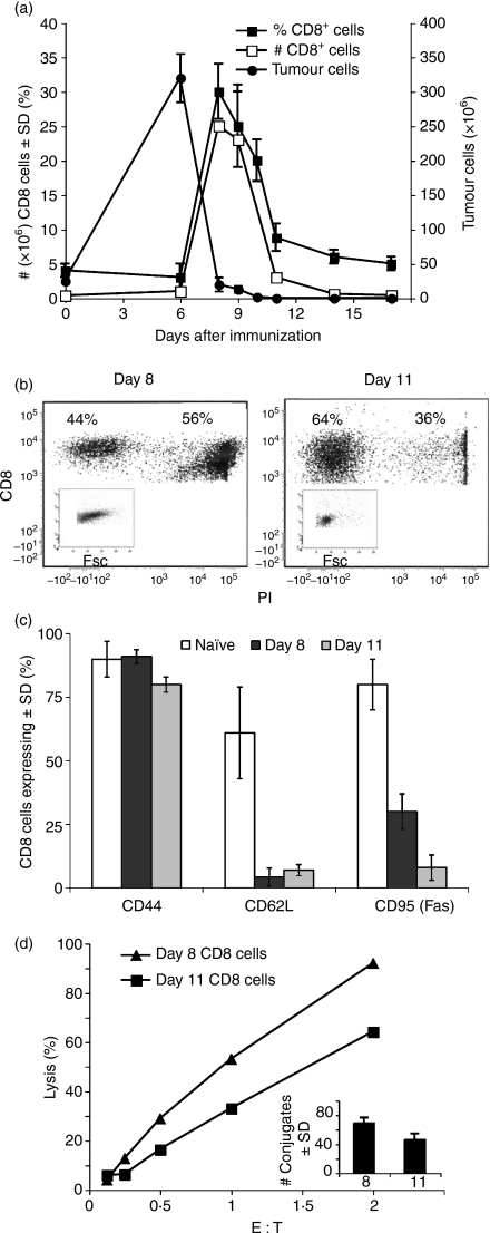
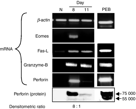
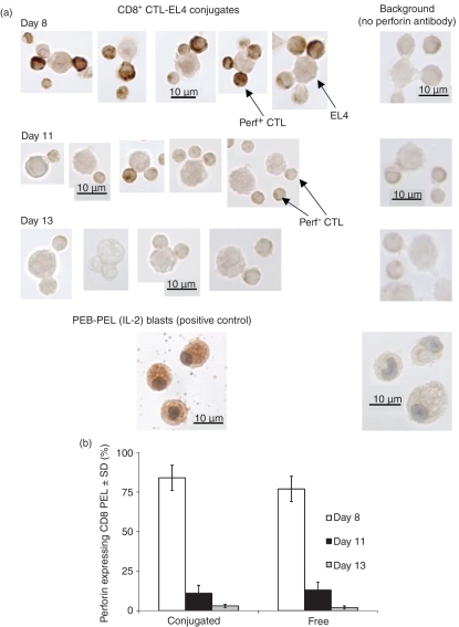
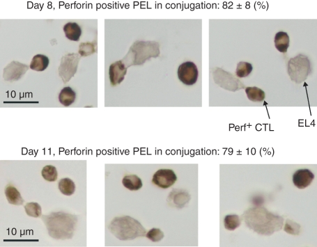
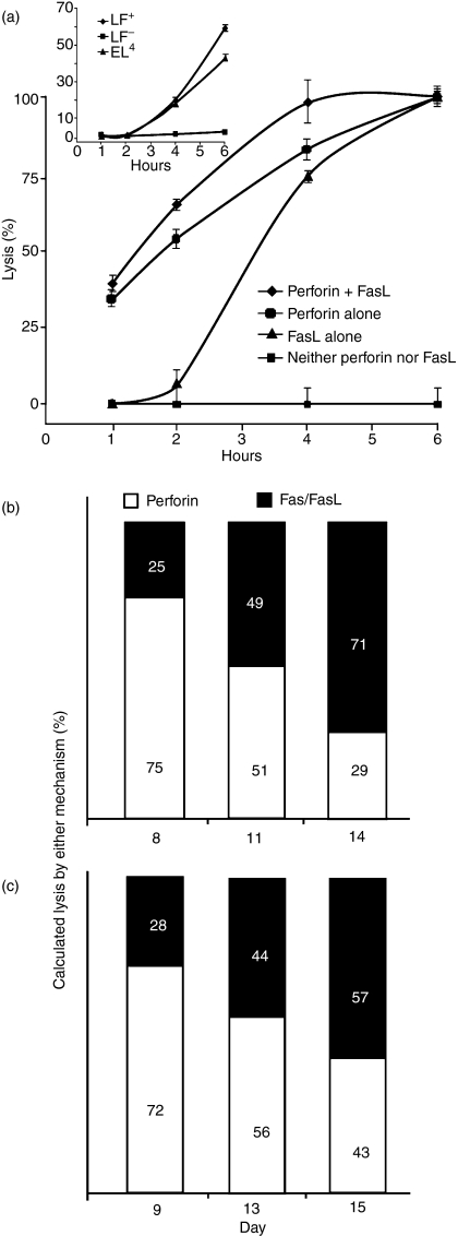
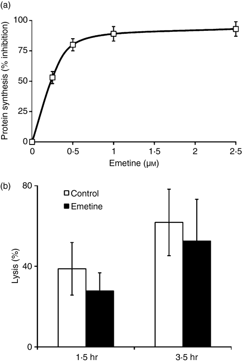
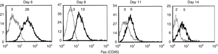
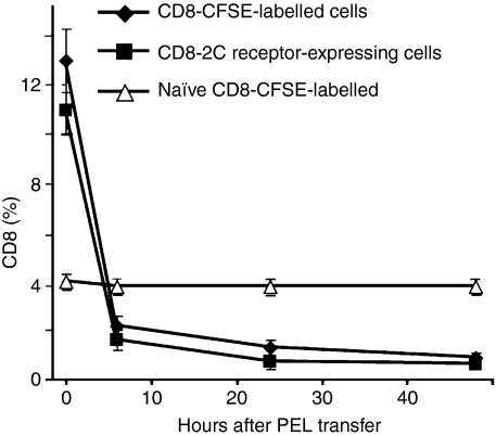
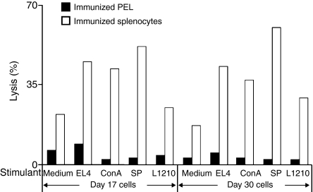
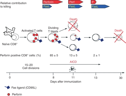
Although CD8(+) cytotoxic T lymphocytes (CTL) exhibit both Fas ligand (FasL) -based and perforin-based lytic activities, the accepted hallmark of a fully active CTL remains its perforin killing machinery. Yet the origin, rationale for possessing both a slow-acting (FasL) and a fast-acting (perforin) killing mechanism has remained enigmatic. Here we have investigated perforin expression in CTL directly involved in acute tumour (i.e. leukaemias EL4 and L1210) allograft rejection occurring within the peritoneal cavity. We show that at the height of the immune response, the majority of conjugate-forming CD8(+) CTL express high levels of perforin messenger RNA and protein, and kill essentially via perforin. Later however, coinciding with complete rejection, fully cytocidal CTL emerge which exhibit a stark decrease in perforin and now kill preferentially via constitutively expressed FasL. Although late in emergence, and persistent, these powerful CTL are neither effector-memory nor memory CTL. This finding has implications for the monitoring of anti-transplant responses in clinical settings, based on assessing perforin expression in graft infiltrating CD8(+) T cells. The results show that as the immune response progresses in vivo, targeted cellular suicide mainly prunes high perforin-expressing CD8(+) cells, resulting in the gradual switch in effector CTL, from mostly perforin-based to largely Fas/FasL-based killers. Hence, two kinds of CD8(+) CTL have two killing strategies.


Figures

Figure 1
Figure 1
CD8+ T-cell response during allograft rejection in the peritoneal cavity. (a) Kinetics of tumour growth and CD8+ T-cell response. BALB/c mice were injected intraperitoneally with allogeneic EL4 tumour cells. Peritoneal exudate lymphocytes (PEL) were, stained for CD8+, the allogeneic tumour for H-2b. Mean ± SD of five animals; one out of three experiments with similar results. (b) CD8 and propidium iodide (PI) staining of responding peritoneal cells. Day 8 and 11 PEL were stained by a CD8+ antibody and analysed by fluorescence-activated cell sorting (FACS) for CD8+ expression. PI staining was used to discern dead cells. Insert depicts forward scatter of CD8+, PI-negative cells. (c) Cell surface markers of responding CD8+ cells. PEL of naïve, day 8 and day 11 immunized mice were stained for CD8+, CD44, CD62L and CD95 (Fas), and analysed by FACS. The mean of four repeat experiments ± SD is shown. (d) Cytotoxicity and conjugate formation by peritoneal CD8+ T cells. Days 8 and 11 immune CD8+ PEL were subjected to a 4-hr cytotoxic assay at different effector-to-target cell (E : T) ratios and % lysis was recorded. In parallel, a fraction of the CD8+ cells was allowed to conjugate at an E : T ratio of 1/1 and the number of conjugates was determined (insert).
Figure 2
Figure 2
Expression of Eomes, Granzyme-B, FasL and perforin in CD8+ T cells. CD8+ peritoneal exudate lymphocytes (PEL) were derived from naive BALB/c mice as well as 8 and 11 days post-injection with allogeneic EL4 tumour cells. Reverse transcription–polymerase chain reaction and Western blot were performed. RNA and protein extracted from PEL blasts (PEB), provided positive controls for FasL, granzyme-B and perforin messenger RNA and protein.
Figure 3
Figure 3
Perforin expression in CD8+ cytotoxic T lymphocytes. Peritoneal exudate lymphocytes (PEL) were extracted from BALB/c anti-EL4 mice 8, 11 or 13 days after tumour injection, CD8+ cells were sorted, excluding propidium iodide-positive cells. (a) CD8+-EL4 conjugates were stained (immunocytochemsitry) using perforin antibody. PEL blasts (PEB) were used as a positive control. Antibody controls were conjugates reacted without the perforin antibody. (b) % Perforin staining of free and conjugated CD8+ sorted cells. Mean ± SD of four repeat experiments, each with cells procured from five animals P(v) > 0·05).
Figure 4
Figure 4
Perforin expression in conjugated CD8+ cytotoxic T lymphocytes obtained after multiple tumour injections. EL4 tumour cells were injected intraperitoenally into BALB/c mice. Eight days later, an additional 25 × 106 EL4 cells were injected daily for 6 days. CD8+ EL4 conjugates were stained (immunocytochemsitry) using perforin antibody.
Figure 5
Figure 5
Differential contribution of the perforin- and Fas-based lytic mechanisms. (a) C57BL/6 and perforin-deficient (PO) anti-LF peritoneal exudate lymphocytes (PEL) were extracted 10 days after immunization and subjected to a 6-hr lytic assay against LF+ and LF cells. Insert: Strictly Fas/FasL-based killing: soluble recombinant FasL trimmer-mediated lysis of LF+ and EL4 commences with a lag of 2 hr; LF cells were not lysed by rFasL. Mean ± SD of three repeat experiments, each with cells procured from three animals. (b) BALB/c anti-EL4 CD8+ PEL subjected to a 1- and 3-hr lytic assay against EL4. Perforin contribution to total killing was calculated: 1 hr/3 hr % lysis; accordingly, FasL-based killing was 100 – (1 hr/3 hr %). One out of three experiments with similar results. (c) C57BL/6 anti-LF CD8+ PEL subjected to a 4-hr lytic assay against LF+ and LF. Lysis of LF was affected by perforin only. Lysis of LF+ was affected by combination of perforin and FasL-based killing. % Perforin killing was calculated as follows: (% LF lysis/% LF+ lysis) × 100, FasL-based killing was calculated: 100 − (% LF lysis/% LF+ lysis) × 100
Figure 6
Figure 6
Emetine effects on protein synthesis and on lysis induced by day 11 peritoneal exudate lymphocytes (PEL). (a) Emetine, 0–2·5 μm, inhibition of protein synthesis in BALB/c anti-EL4 PEL. (b) Inhibition of lysis by day 11 PEL pre-incubated with Emetine (2·5 μm, for 2 hr) before a 1·5-hr or 3·45-hr lytic assay in the presence of Emetine; effector-to-target ratio of 10 : 1. Mean ± SD of three repeat experiments, each with cells procured from three animals is shown. P(v) < 0·05).
Figure 7
Figure 7
Fas expression on responding CD8+ peritoneal exudate lymphocytes (PEL). Mean fluorescence intensity (MFI, in italics) of Fas+ CD8+ PEL extracted from the peritoneal cavity of C57BL/6 anti-LF mice 6, 8, 11 and 14 days after immunization. Animals were killed and the cells were analysed on the same day. Cells were stained with CD8+ and Fas (CD95) antibodies and analysed by fluorescence-activated cell sorting. The grey line depicts cells stained for CD8+ only and serve as a control. One of three experiments with similar though variable patterns is shown.
Figure 8
Figure 8
The fate of day 8 CD8+ peritoneal exudate lymphocytes (PEL) in vivo. Day 8 BALB/c anti-EL4 PEL were stained with CFSE, analysed for CD8+ CFSE+ cells, and then reinjected intraperitoneally (i.p.) into remaining mice. Parallel experiments were conducted with immunized transgenic 2C and C57BL/6 anti-LF mice. PEL from the 2C mice were extracted 8 or 11 days after immunization and reinjected i.p. into C57BL/6 mice of the same day, i.e. day 8 PEL were injected into day 8 mice. PEL were removed from the latter mice at different times and the CD8+/CFSE+ or 2C CD8+ population was analysed by fluorescence-activated cell sorting. Each experiment was performed twice.
Figure 9
Figure 9
Restimulation of primed splenocytes and peritoneal exudate lymphocytes (PEL). PEL and splenocytes were collected from BALB/c anti-EL4 mice injected 17 and 30 days before were subjected to a 3-day restimulation with LF+, EL4, irradiated C57BL/6 splenocytes, concanavalin A (2 μg/ml), and with medium as control. Cells were then tested in a 4-hr lytic assay against EL4 and % lysis was recorded. One out of three experiments performed is illustrated.
Figure 10
Figure 10
The transition from mostly perforin to FasL cytotoxic T lymphocytes (CTL). The CTL start off as naïve CD8+ T cells, which upon activation by antigen-laden antigen-presenting cells, and cytokines develop into dividing CTL blasts that continue to proliferate and differentiate into effector CTL of decreasing sizes. Initially, the responding CTL possess both the perforin and FasL killing mechanisms. As the immune reaction progresses, and antigen becomes limiting, most CTL blasts are pruned (probably by activation-induced cell death); the remaining CTL, now of decreasing sizes, gradually stop expressing perforin, relying mainly on FasL activity. These CTL are not memory cells however.

Similar articles

Cited by

References

    1. Cerottini JC, Brunner KT. Cell-mediated cytotoxicity, allograft rejection, and tumor immunity. Adv Immunol. 1974;18:67–132. - PubMed
    1. Kägi D, Ledermann B, Burki K, Zinkernagel RM, Hengartner H. Molecular mechanisms of lymphocyte-mediated cytotoxicity and their role in immunological protection and pathogenesis in vivo. Annu Rev Immunol. 1996;14:207–32. - PubMed
    1. Trapani JA, Smyth MJ. Functional significance of the perforin/granzyme cell death pathway. Nat Rev Immunol. 2002;2:735–47. - PubMed
    1. Henkart PA, Catalfamo M. CD8+ effector cells. Adv Immunol. 2004;83:233–52. - PubMed
    1. Berke G, Clark W. Killer lymphocytes. Berlin: Springer; 2005.

Publication types