. 2009 Sep;128(1):141-9.
doi: 10.1111/j.1365-2567.2009.03105.x.

Increase in frequency of myeloid-derived suppressor cells in mice with spontaneous pancreatic carcinoma

Affiliations

Increase in frequency of myeloid-derived suppressor cells in mice with spontaneous pancreatic carcinoma

Fei Zhao et al. Immunology. 2009 Sep.

Abstract

Pancreatic adenocarcinoma is one of the deadliest cancers with poor survival and limited treatment options. Immunotherapy is an attractive option for this cancer that needs to be further developed. Tumours have evolved a variety of mechanisms to suppress host immune responses. Understanding these responses is central in developing immunotherapy protocols. The aim of this study was to investigate potential immune suppressor mechanisms that might occur during development of pancreatic tumours. Myeloid-derived suppressor cells (MDSC) from mice with spontaneous pancreatic tumours, mice with premalignant lesions as well as wild-type mice were analysed. An increase in the frequency of MDSC early in tumour development was detected in lymph nodes, blood and pancreas of mice with premalignant lesions and increased further upon tumour progression. The MDSC from mice with pancreatic tumours have arginase activity and suppress T-cell responses, which represent the hallmark functions of these cells. Our study suggests that immune suppressor mechanisms generated by tumours exist as early as premalignant lesions and increase with tumour progression. These results highlight the importance of blocking these suppressor mechanisms early in the disease in developing immunotherapy protocols.


Figures

Figure 1
Figure 1
Histological analysis of pancreas from elastase-transforming growth factor-α (EL-TGF-α)/p53−/− (a), EL-TGF-α/p53+/− (b) and wild-type (c) mice.
Figure 2
Figure 2
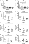
Frequency of myeloid-derived suppressor cells (MDSC) in elastase-transforming growth factor-α (EL-TGF-α)/p53−/− tumour-bearing mice, EL-TGF-α/p53+/−, mice with premalignant lesions, age-matched wild-type mice and mPAC challenged wild-type mice (mPAC). The frequency of MDSC was determined in spleen (a), blood (b), mesenteric lymph nodes (c) and pancreas (d) and is represented as the percentage of CD11b+ Gr-1+ cells in the monocyte and lymphocyte populations. CD11b+ Gr-1high and CD11b+ Gr-1dull MDSC subtypes were analysed in spleen (e, f) and blood (g, h) of mice with established tumours, premalignant lesions and wild-type mice. (*) indicates a P-value of < 0·05.
Figure 3
Figure 3
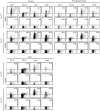
Phenotypic analysis of Gr-1high and Gr-1dull myeloid-derived suppressor cells (MDSC) derived from mice with pancreatic tumours, premalignant lesions and subcutaneous mPAC tumours. CD11b+ Gr-1high and Gr-1dull MDSC were gated and analysed for the indicated markers. The isotype control is shown for every staining.
Figure 4
Figure 4
Arginase activity of myeloid-derived suppressor cells (MDSC) from mice with established pancreatic tumours, mice with premalignant pancreatic lesions and wild-type mice. Data shown represent the mean value of four independent experiments. (*) indicates a P-value of < 0·05.
Figure 6
Figure 6
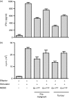
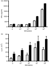
Analysis of the suppressive function of CD11b+ Gr-1high and CD11b+ Gr-1dull myeloid-derived suppressor cells (MDSC) derived from mice with established pancreatic tumours and premalignant lesions: FACS-sorted CD11b+ Gr-1high or CD11b+ Gr-1dull MDSC from mice with premalignant lesions or established tumours were added to C57BL/6 splenocytes stimulated with irradiated allogeneic splenocytes. (a) IFN-γ secretion and (b) proliferation of T cells was measured in cell supernatants and by thymidine incorporation after 48 hr. Data shown represent the average of four mice.
Figure 5
Figure 5
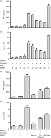
Analysis of the suppressive function of myeloid-derived suppressor cells (MDSC) derived from mice with established pancreatic tumours: (a, b) Increasing numbers of fluorescence-activated cell sorting (FACS)-analysed MDSC from elastase-transforming growth factor-α (EL-TGF-α)/p53−/− mice were added to C57BL/6 splenocytes stimulated with irradiated allogeneic splenocytes. Interferon-γ (IFN-γ) secretion and proliferation of T cells was measured after 48 hr in cell supernatants and by thymidine incorporation. Data shown represent one of four independent experiments with similar results. (c, d) FACS-sorted MDSC from mice with premalignant lesions, established tumours and naive mice were added to C57BL/6 splenocytes stimulated with irradiated allogeneic splenocytes. IFN-γ secretion and proliferation of T cells was measured in cell supernatants and by thymidine incorporation after 48 hr. Data shown represent one of four independent experiments with similar results.
Figure 7
Figure 7
Depletion of myeloid-derived suppressor cells (MDSC) from splenocytes isolated from mice with established tumours enhances T-cell responses: (a) C57BL/6 splenocytes were stimulated with increasing numbers of allogeneic splenocytes derived from tumour-bearing mice (white bars) or after depletion (black bars) of MDSC. Interferon-γ (IFN-γ) secretion was determined in supernatants after 48 hr. Data represent one of four independent experiments with similar results. (b) BALB/c splenocytes were stimulated with increasing numbers of mitomycin-C-treated allogeneic splenocytes derived from tumour-bearing mice (white bars) or after depletion (black bars) of MDSC. Proliferation was measured by thymidine incorporation after 3 days. Data represent one of four independent experiments with similar results.

References

    1. Jemal A, Siegel R, Ward E, Hao Y, Xu J, Murray T, Thun MJ. Cancer Statistics, 2008. CA Cancer J Clin. 2008;58:71–96. - PubMed
    1. Laheru D, Jaffee EM. Immunotherapy for pancreatic cancer – science driving clinical progress. Nat Rev. 2005;5:459–67. - PubMed
    1. Garbe AI, Vermeer B, Gamrekelashvili J, et al. Genetically induced pancreatic adenocarcinoma is highly immunogenic and causes spontaneous tumor-specific immune responses. Cancer Res. 2006;66:508–16. - PubMed
    1. Wagner M, Greten FR, Weber CK, et al. A murine tumor progression model for pancreatic cancer recapitulating the genetic alterations of the human disease. Genes Dev. 2001;15:286–93. - PMC - PubMed
    1. Wagner M, Luhrs H, Kloppel G, Adler G, Schmid RM. Malignant transformation of duct-like cells originating from acini in transforming growth factor transgenic mice. Gastroenterology. 1998;115:1254–62. - PubMed

Publication types

MeSH terms