The added value of longitudinal black-blood cardiovascular magnetic resonance angiography in the cross sectional identification of carotid atherosclerotic ulceration
- PMID: 19689816
- PMCID: PMC2737539
- DOI: 10.1186/1532-429X-11-31
The added value of longitudinal black-blood cardiovascular magnetic resonance angiography in the cross sectional identification of carotid atherosclerotic ulceration
Abstract
Background: Carotid atherosclerotic ulceration is a significant source of stroke. This study evaluates the efficacy of adding longitudinal black-blood (BB) cardiovascular magnetic resonance (CMR) angiography to cross-sectional CMR images in the identification of carotid atherosclerotic ulceration.
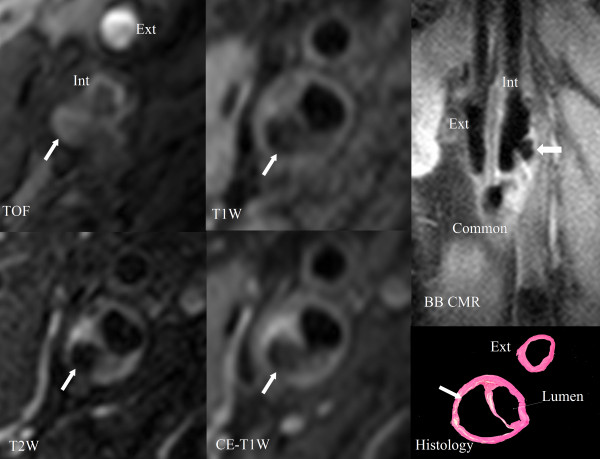
Methods: Thirty-two subjects (30 males and two females with ages between 48 and 83 years) scheduled for carotid endarterectomy were imaged on a 1.5T GE Signa scanner using multisequence [3D time-of-flight, T1, proton density, T2, contrast enhanced T1], cross-sectional CMR images and longitudinal BB CMR angiography (0.625 x 0.625 mm/pixel). Two rounds of review (round 1: cross-sectional CMR images alone and round 2: cross-sectional CMR images plus longitudinal BB CMR angiography) were conducted for the presence and volume measurements of ulceration. Ulceration was defined as a distinct depression into the plaque containing blood flow signal on cross-sectional CMR and longitudinal BB CMR angiography.
Results: Of the 32 plaques examined by histology, 17 contained 21 ulcers. Using the longitudinal BB CMR angiography sequence in addition to the cross-sectional CMR images in round 2, the sensitivity improved to 80% for ulcers of at least 6 mm3 in volume by histology and 52.4% for all ulcers, compared to 30% and 23.8% in round 1, respectively. There was a slight decline in specificity from 88.2% to 82.3%, though both the positive and negative predictive values increased modestly from 71.4% to 78.6% and from 48.4% to 58.3%, respectively.
Conclusion: The addition of longitudinal BB CMR angiography to multisequence cross-sectional CMR images increases accuracy in the identification of carotid atherosclerotic ulceration.
Figures


Similar articles
-
Computed tomography angiography vs 3 T black-blood cardiovascular magnetic resonance for identification of symptomatic carotid plaques.J Cardiovasc Magn Reson. 2014 Oct 7;16(1):84. doi: 10.1186/s12968-014-0084-y. J Cardiovasc Magn Reson. 2014. PMID: 25315518 Free PMC article.
-
High signal intensity halo around the carotid artery on maximum intensity projection images of time-of-flight MR angiography: a new sign for intraplaque hemorrhage.J Magn Reson Imaging. 2008 Jun;27(6):1341-6. doi: 10.1002/jmri.21284. J Magn Reson Imaging. 2008. PMID: 18504753 Clinical Trial.
-
Comparison of symptomatic and asymptomatic atherosclerotic carotid plaques using parallel imaging and 3 T black-blood in vivo CMR.J Cardiovasc Magn Reson. 2013 May 24;15(1):44. doi: 10.1186/1532-429X-15-44. J Cardiovasc Magn Reson. 2013. PMID: 23705576 Free PMC article.
-
Evaluation of carotid artery atherosclerotic plaque distribution by using long-axis high-resolution black-blood magnetic resonance imaging.J Neurosurg. 2008 Dec;109(6):1042-8. doi: 10.3171/JNS.2008.109.12.1042. J Neurosurg. 2008. PMID: 19035717
-
Carotid plaque regression following 6-month statin therapy assessed by 3T cardiovascular magnetic resonance: comparison with ultrasound intima media thickness.J Cardiovasc Magn Reson. 2011 Aug 3;13(1):37. doi: 10.1186/1532-429X-13-37. J Cardiovasc Magn Reson. 2011. PMID: 21812992 Free PMC article.
Cited by
-
Intraplaque Hemorrhage and the Plaque Surface in Carotid Atherosclerosis: The Plaque At RISK Study (PARISK).AJNR Am J Neuroradiol. 2015 Nov;36(11):2127-33. doi: 10.3174/ajnr.A4414. Epub 2015 Aug 6. AJNR Am J Neuroradiol. 2015. PMID: 26251429 Free PMC article. Clinical Trial.
-
Prediction of high-risk plaque development and plaque progression with the carotid atherosclerosis score.JACC Cardiovasc Imaging. 2014 Apr;7(4):366-73. doi: 10.1016/j.jcmg.2013.09.022. Epub 2014 Mar 13. JACC Cardiovasc Imaging. 2014. PMID: 24631510 Free PMC article.
-
Magnetic resonance imaging of carotid plaques: current status and clinical perspectives.Ann Transl Med. 2020 Oct;8(19):1266. doi: 10.21037/atm-2020-cass-16. Ann Transl Med. 2020. PMID: 33178798 Free PMC article. Review.
-
Diagnostics Accuracy of Magnetic Resonance Imaging in Detection of Atherosclerotic Plaque Characteristics in Carotid Arteries Compared to Histology: A Systematic Review.J Magn Reson Imaging. 2025 Mar;61(3):1067-1093. doi: 10.1002/jmri.29522. Epub 2024 Jul 9. J Magn Reson Imaging. 2025. PMID: 38981139 Free PMC article.
-
Repeatability of in vivo quantification of atherosclerotic carotid artery plaque components by supervised multispectral classification.MAGMA. 2015 Dec;28(6):535-45. doi: 10.1007/s10334-015-0495-2. Epub 2015 Jul 11. MAGMA. 2015. PMID: 26162931 Free PMC article.
References
-
- Rosamond W, Flegal K, Furie K, Go A, Greenlund K, Haase N, Hailpern SM, Ho M, Howard V, Kissela B, Kittner S, Lloyd-Jones D, McDermott M, Meigs J, Moy C, Nichol G, O'Donnell C, Roger V, Sorlie P, Steinberger J, Thom T, Wilson M, Hong Y. Heart disease and stroke statistics 2008 update: a report from the American Heart Association Statistics Committee and Stroke Statistics Subcommittee. Circulation. 2008;117:e25–e146. doi: 10.1161/CIRCULATIONAHA.107.187998. - DOI - PubMed
Publication types
MeSH terms
Substances
Grants and funding
LinkOut - more resources
Full Text Sources
Medical