. 2009 Oct;1792(10):1036-42.
doi: 10.1016/j.bbadis.2009.08.002. Epub 2009 Aug 16.

Ribosomal protein S19 and S24 insufficiency cause distinct cell cycle defects in Diamond-Blackfan anemia

Affiliations

Ribosomal protein S19 and S24 insufficiency cause distinct cell cycle defects in Diamond-Blackfan anemia

Jitendra Badhai et al. Biochim Biophys Acta. 2009 Oct.

Abstract

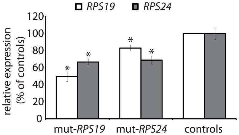
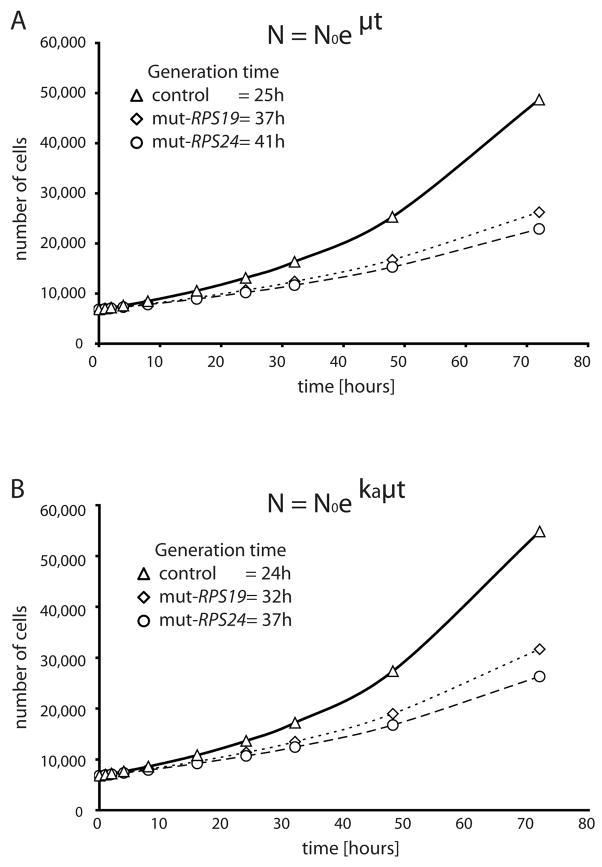
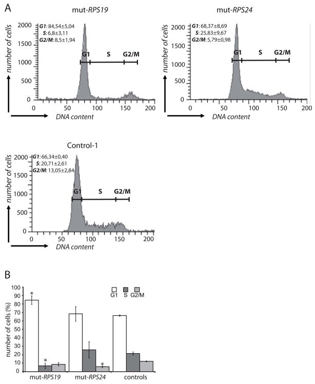
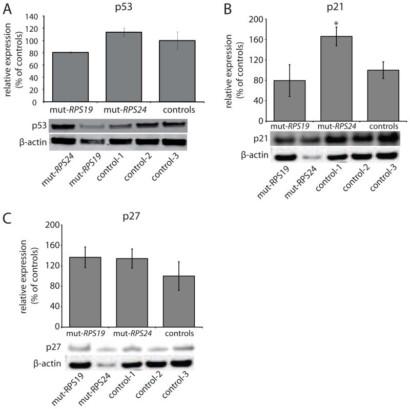
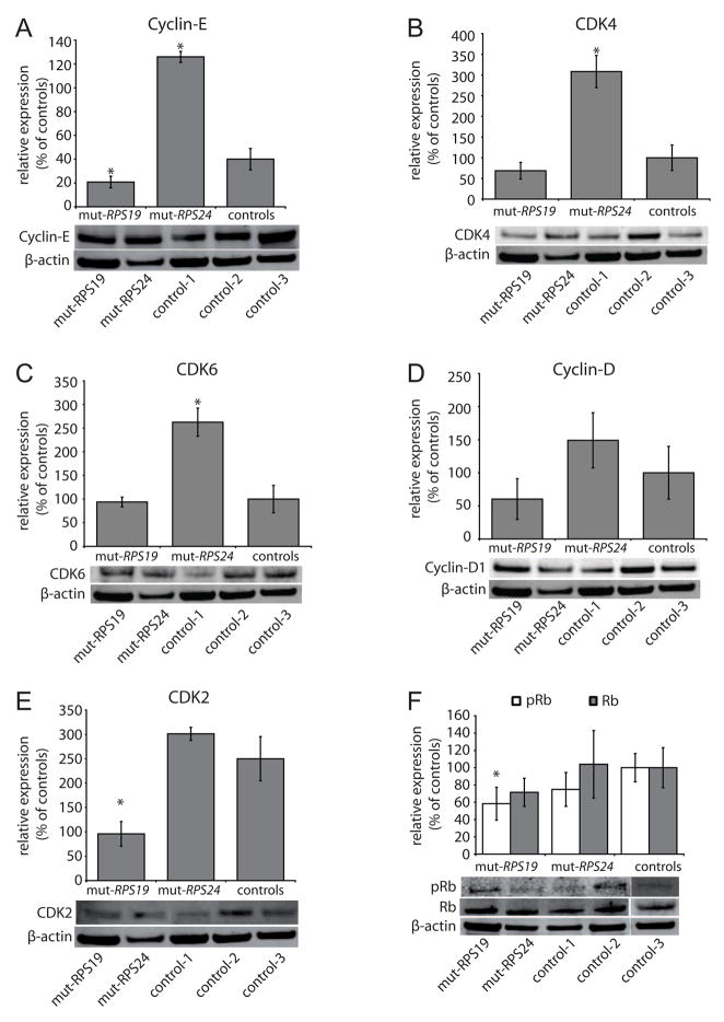
Diamond-Blackfan anemia (DBA) is a severe congenital anemia characterized by a specific decrease of erythroid precursors. The disease is also associated with growth retardation, congenital malformations, a predisposition for malignant disease and heterozygous mutations in either of the ribosomal protein (RP) genes RPS7, RPS17, RPS19, RPS24, RPL5, RPL11 and RPL35a. We show herein that primary fibroblasts from DBA patients with truncating mutations in RPS19 or in RPS24 have a marked reduction in proliferative capacity. Mutant fibroblasts are associated with extended cell cycles and normal levels of p53 when compared to w.t. cells. RPS19 mutant fibroblasts accumulate in the G1 phase, whereas the RPS24 mutant cells show an altered progression in the S phase resulting in reduced levels in the G2/M phase. RPS19 deficient cells exhibit reduced levels of Cyclin-E, CDK2 and retinoblastoma (Rb) protein supporting a cell cycle arrest in the G1 phase. In contrast, RPS24 deficient cells show increased levels of the cell cycle inhibitor p21 and a seemingly opposing increase in Cyclin-E, CDK4 and CDK6. In combination, our results show that RPS19 and RPS24 insufficient fibroblasts have an impaired growth caused by distinct blockages in the cell cycle. We suggest this proliferative constraint to be an important contributing mechanism for the complex extra-hematological features observed in DBA.


Figures

Figure 1
Figure 1
Levels of RPS19 and RPS24 proteins in primary fibroblasts as determined by western blot. Relative levels of RPS19 protein (white bar) and RPS24 protein (grey bar) in RPS19 (mut-RPS19) and RPS24 (mut-RPS24) mutant cells as compared to controls (100% expression). Asterix (*) indicates significant differences when compared to control cells (p<0.05).
Figure 2
Figure 2
Growth curves of primary fibroblasts carrying RPS19 and RPS24 mutations, respectively, and three control fibroblasts. (A) 6,800 cells/cm2 were seeded in duplicates and counted after 48 h and 72 h to measure the generation time (tgen). A growth curve assuming exponential growth is simulated using equation N= Noeμt showing the number of cells as a function of time. μ= 0,693/tgen. (B) Replotted growth curves from (A) implementing an apoptosis correction factor (ka) to obtain the adjusted growth curve equation N=Noekaμt. RPS19 mutant cells (open squares), RPS24 mutant cells (open circles) and control fibroblasts (open triangles).
Figure 3
Figure 3
Cell cycle analysis of human primary fibroblasts. The cell cycle phases were measured by propidium iodide labeling. (A) Representative histograms from cell cycle analysis of the RPS19 mutated (mut-RPS19), RPS24 mutated (mut-RPS24) cells and three controls. The cell cycle phases G1, S and G2/M are indicated. (B) Diagram showing the distribution of cells in distinct cell cycle phases from two independent experiments. The average percentages of cells in the different cell cycle phases (G1, S and G2/M) are shown with standard deviations. Asterix (*) indicates significant differences when compared to control cells in the same phase, respectively (p<0.05).
Figure 4
Figure 4
Analysis of p53, p21 and p27 protein levels in fibroblasts carrying RPS19 mutation (mut-RPS19), RPS24 mutation (mut-RPS24) and controls (100% expression). Western blot analysis were normalised to beta-actin. (A) p53 protein expression. The diagram illustrates the average of two independent measurements and standard deviations without significant differences. Representative analysis (bottom) using antibodies against p53 and β-actin, respectively. (B) Relative levels of p21 in RPS19 and RPS24 mutant cells from two different experiments and controls (100% expression). Asterix (*) denotes significant differences compared to controls (p<0.05). Representative analysis (bottom) using antibodies against p21 and β-actin, respectively. (C) Relative levels of p27 in RPS19 and RPS24 mutant cells as compared to controls (100% expression). Representative analysis (bottom) using antibodies against p27 and β-actin, respectively.
Figure 5
Figure 5
Western blot analysis of the cell cycle regulators Cyclin-E, CDK4, CDK6, Cyclin-D1, CDK2 and retinoblastoma protein (Rb) in fibroblasts carrying RPS19 mutation (mut-RPS19), RPS24 mutation (mut-RPS24) and three controls (100% expression). Averages of at least two independent experiments are shown with standard deviations. The western analysis was normalized to β-actin and representative blots are illustrated below each diagram. Asterix (*) denotes significant differences (p<0.05) compared to controls. (A) Relative levels of Cyclin-E. (B) Relative levels of CDK4. (C) Relative levels of CDK6. (D) Relative levels of Cyclin-D showing tendencies for variations in RPS19 and RPS24 mutant cells but these are non-significant. (E) Relative levels of CDK2. (F) Relative levels of phosphorylated Rb (pRb; white bars) and non-phosphorylated Rb (Rb; grey bar) proteins.

References

    1. Young NS, Alter B. Inherited bone marrow failure syndromes: Introduction. In: Young, Alter, editors. Aplastic anemia acquired and inherited. WB saunders Campany; Philadelphia, PA: 1994.
    1. Halperin DS, Freedman MH. Diamond-blackfan anemia: etiology, pathophysiology, and treatment. Am J Pediatr Hematol Oncol. 1989;11:380–394. - PubMed
    1. Willig TN, Draptchinskaia N, Dianzani I, Ball S, Niemeyer C, Ramenghi U, Orfali K, Gustavsson P, Garelli E, Brusco A, Tiemann C, Perignon JL, Bouchier C, Cicchiello L, Dahl N, Mohandas N, Tchernia G. Mutations in ribosomal protein S19 gene and diamond blackfan anemia: wide variations in phenotypic expression. Blood. 1999;94:4294–4306. - PubMed
    1. Gripp KW, McDonald-McGinn DM, La Rossa D, McGain D, Federman N, Vlachos A, Glader BE, McKenzie SE, Lipton JM, Zackai EH. Bilateral microtia and cleft palate in cousins with Diamond-Blackfan anemia. Am J Med Genet. 2001;101:268–274. - PubMed
    1. Gazda HT, Sheen MR, Vlachos A, Choesmel V, O’Donohue MF, Schneider H, Darras N, Hasman C, Sieff CA, Newburger PE, Ball SE, Niewiadomska E, Matysiak M, Zaucha JM, Glader B, Niemeyer C, Meerpohl JJ, Atsidaftos E, Lipton JM, Gleizes PE, Beggs AH. Ribosomal protein L5 and L11 mutations are associated with cleft palate and abnormal thumbs in Diamond-Blackfan anemia patients. Am J Hum Genet. 2008;83:769–780. - PMC - PubMed

Publication types