

A 2-helix small protein labeled with 68Ga for PET imaging of HER2 expression
- PMID: 19690041
- PMCID: PMC4216181
- DOI: 10.2967/jnumed.109.064287
A 2-helix small protein labeled with 68Ga for PET imaging of HER2 expression
Abstract
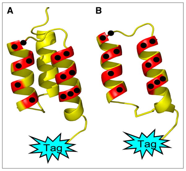
Affibody molecules are a class of scaffold proteins being developed into a generalizable approach to targeting tumors. Many 3-helix-based Affibody proteins have shown excellent in vivo properties for tumor imaging and therapy. By truncating one alpha-helix that is not responsible for receptor recognition in the Affibody and maturating the protein affinity through synthetic strategies, we have successfully identified in our previous research several small 2-helix proteins with excellent binding affinities to human epidermal growth factor receptor type 2 (HER2). With preferential properties such as faster blood clearance and tumor accumulation, lower immunogenic potential, and facile and economically viable synthetic schemes, we hypothesized that these 2-helix protein binders could become excellent molecular imaging probes for monitoring HER2 expression and modulation.
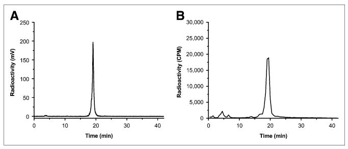
Methods: In this study, a 2-helix small protein, MUT-DS, was chemically modified with a metal chelator, 1,4,7,10-tetraazacyclododecane-1,4,7,10-tetraacetic acid (DOTA). DOTA-MUT-DS was then site-specifically radiolabeled with an important PET radionuclide, (68)Ga. The resulting radiolabeled anti-HER2 2-helix molecule was further evaluated as a potential molecular probe for small-animal PET HER2 imaging in a SKOV3 tumor mouse model.
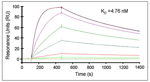
Results: The 2-helix DOTA-MUT-DS showed high HER2-binding affinity (dissociation constant, 4.76 nM). The radiolabeled probe displayed high stability in mouse serum and specificity toward HER2 in cell cultures. Biodistribution and small-animal PET studies further showed that (68)Ga-DOTA-MUT-DS had rapid and high SKOV3 tumor accumulation and quick clearance from normal organs. The specificity of (68)Ga-DOTA-MUT-DS for SKOV3 tumors was confirmed by monitoring modulation of HER2 protein on treatment of tumor mice with heat shock protein 90 inhibitor 17-N,N-dimethyl ethylene diamine-geldanamycin in vivo.
Conclusion: This proof-of-concept research clearly demonstrated that synthetic 2-helix (68)Ga-DOTA-MUT-DS is a promising PET probe for imaging HER2 expression in vivo. The Affibody-derived small 2-helix protein scaffold has great potential for developing targeting agents for a variety of tumor-associated biomarkers.
Figures


References
-
- Nygren PA. Alternative binding proteins: affibody binding proteins developed from a small three-helix bundle scaffold. FEBS J. 2008;275:2668–2676. - PubMed
-
- Nygren PA, Skerra A. Binding proteins from alternative scaffolds. J Immunol Methods. 2004;290:3–28. - PubMed
-
- Orlova A, Magnusson M, Eriksson TL, et al. Tumor imaging using a picomolar affinity HER2 binding affibody molecule. Cancer Res. 2006;66:4339–4348. - PubMed
-
- Orlova A, Tolmachev V, Pehrson R, et al. Synthetic affibody molecules: a novel class of affinity ligands for molecular imaging of HER2-expressing malignant tumors. Cancer Res. 2007;67:2178–2186. - PubMed
-
- Nordberg E, Orlova A, Friedman M, et al. In vivo and in vitro uptake of 111In, delivered with the affibody molecule (ZEGFR:955)2, in EGFR expressing tumour cells. Oncol Rep. 2008;19:853–857. - PubMed
Publication types
MeSH terms
Substances
Grants and funding
LinkOut - more resources
Full Text Sources
Other Literature Sources
Medical
Research Materials
Miscellaneous