

Will the original glucose transporter isoform please stand up!
- PMID: 19690067
- PMCID: PMC2763785
- DOI: 10.1152/ajpendo.00496.2009
Will the original glucose transporter isoform please stand up!
Abstract
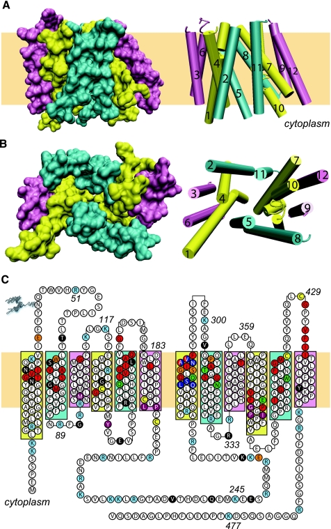
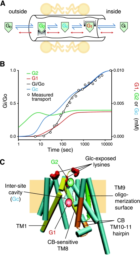
Monosaccharides enter cells by slow translipid bilayer diffusion by rapid, protein-mediated, cation-dependent cotransport and by rapid, protein-mediated equilibrative transport. This review addresses protein-mediated, equilibrative glucose transport catalyzed by GLUT1, the first equilibrative glucose transporter to be identified, purified, and cloned. GLUT1 is a polytopic, membrane-spanning protein that is one of 13 members of the human equilibrative glucose transport protein family. We review GLUT1 catalytic and ligand-binding properties and interpret these behaviors in the context of several putative mechanisms for protein-mediated transport. We conclude that no single model satisfactorily explains GLUT1 behavior. We then review GLUT1 topology, subunit architecture, and oligomeric structure and examine a new model for sugar transport that combines structural and kinetic analyses to satisfactorily reproduce GLUT1 behavior in human erythrocytes. We next review GLUT1 cell biology and the transcriptional and posttranscriptional regulation of GLUT1 expression in the context of development and in response to glucose perturbations and hypoxia in blood-tissue barriers. Emphasis is placed on transgenic GLUT1 overexpression and null mutant model systems, the latter serving as surrogates for the human GLUT1 deficiency syndrome. Finally, we review the role of GLUT1 in the absence or deficiency of a related isoform, GLUT3, toward establishing the physiological significance of coordination between these two isoforms.
Figures
References
-
- Abramson J, Smirnova I, Kasho V, Verner G, Kaback HR, Iwata S. Structure and mechanism of the lactose permease of Escherichia coli. Science 301: 610–615, 2003 - PubMed
-
- Alvarez J, Lee DC, Baldwin SA, Chapman D. Fourier transform infrared spectroscopic study of the structure and conformational changes of the human erythrocyte glucose transporter. J Biol Chem 262: 3502–3509, 1987 - PubMed
-
- Appleman JR, Lienhard GE. Kinetics of the purified glucose transporter. Direct measurement of the rates of interconversion of transporter conformers. Biochemistry 28: 8221–8227, 1989 - PubMed
-
- Badr GA, Tang J, Ismail-Beigi F, Kern TS. Diabetes downregulates Glut1 expression in the retina and its microvessels but not in the cerebral cortex or its microvessels. Diabetes 49: 1016–1021, 2000 - PubMed
-
- Baker GF, Naftalin RJ. Evidence of multiple operational affinities for d-glucose inside the human erythrocyte membrane. Biochim Biophys Acta 550: 474–484, 1979 - PubMed
Publication types
MeSH terms
Substances
Grants and funding
LinkOut - more resources
Full Text Sources
Miscellaneous