. 2009 Sep 1;69(17):6889-98.
doi: 10.1158/0008-5472.CAN-09-0347. Epub 2009 Aug 18.

Fyn and SRC are effectors of oncogenic epidermal growth factor receptor signaling in glioblastoma patients

Affiliations

Fyn and SRC are effectors of oncogenic epidermal growth factor receptor signaling in glioblastoma patients

Kan V Lu et al. Cancer Res. .

Abstract

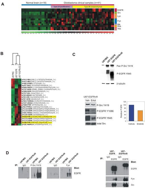
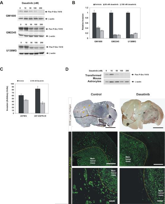
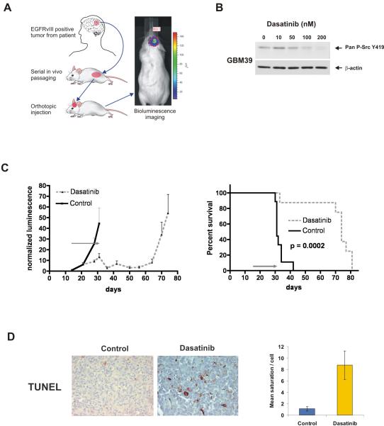
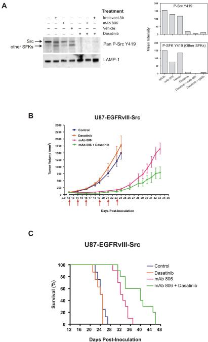
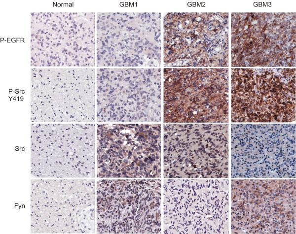
Activating epidermal growth factor receptor (EGFR) mutations are common in many cancers including glioblastoma. However, clinical responses to EGFR inhibitors are infrequent and short-lived. We show that the Src family kinases (SFK) Fyn and Src are effectors of oncogenic EGFR signaling, enhancing invasion and tumor cell survival in vivo. Expression of a constitutively active EGFR mutant, EGFRvIII, resulted in activating phosphorylation and physical association with Src and Fyn, promoting tumor growth and motility. Gene silencing of Fyn and Src limited EGFR- and EGFRvIII-dependent tumor cell motility. The SFK inhibitor dasatinib inhibited invasion, promoted tumor regression, and induced apoptosis in vivo, significantly prolonging survival of an orthotopic glioblastoma model expressing endogenous EGFRvIII. Dasatinib enhanced the efficacy of an anti-EGFR monoclonal antibody (mAb 806) in vivo, further limiting tumor growth and extending survival. Examination of a large cohort of clinical samples showed frequent coactivation of EGFR and SFKs in glioblastoma patients. These results establish a mechanism linking EGFR signaling with Fyn and Src activation to promote tumor progression and invasion in vivo and provide rationale for combined anti-EGFR and anti-SFK targeted therapies.


Figures

Figure 1
Figure 1
SFKs are expressed in glioblastomas and are direct effectors of oncogenic EGFR signaling. A, Microarray analysis shows Fyn, Src, and Yes expression across all glioblastomas. Fyn expression was positively correlated with EGFR expression (r2=0.62, p<0.001). B, Global phosphotyrosine profiling identified Y420 and Y185 Fyn phosphorylation in association with EGFR/EGFRvIII overexpression in EGF-stimulated U87 cells. C, Western blot analysis confirmed upregulation of SFK Y419 phosphorylation and EGFR Y845 phosphorylation, a Src substrate site, in EGFR/EGFRvIII-expressing U87 cells (top). Erlotinib inhibits EGFR Y1086 autophosphorylation and mitigates SFK Y419 and EGFR Y845 phosphorylation mediated by EGFRvIII (bottom left). Quantification of P-Src Y419 inhibition due to erlotinib treatment (bottom right). D, U87MG or U87-EGFRvIII lysates immunoprecipitated with pan-P-Src Y419 or Fyn antibodies were immunoblotted to detect EGFR, demonstrating physical interaction between Fyn/phospho-SFKs and EGFRvIII (left). Right, U87-EGFR and U87-EGFRvIII lysates pulled down with EGFR antibodies were immunoblotted against EGFR, Fyn, and Src to demonstrate physical interaction between Fyn /Src and EGFR.
Figure 2
Figure 2
Fyn and Src promote motility of EGFR-expressing glioblastoma cells. A, Primary glioblastoma cultures and established glioblastoma cell lines expressing EGFR exhibit high levels of activating SFK phosphorylation. U87MG cells express low levels of EGFR and have relatively low phospho-SFK activation. B, Glioblastoma cell lines transfected with Fyn-specific siRNAs (Fyn) demonstrated 50-90% reduction in tumor cell invasion through Matrigel compared to control scrambled siRNA (Sc) (mean P=0.017). U87MG cells transfected with Fyn cDNA were more invasive in vitro. C, EGFRvIII expression promotes SFK phosphorylation and U87 cell invasion through Matrigel. Constitutively active Src significantly enhances invasion and total SFK phosphorylation; an EGFRvIII Y845F mutant suppresses invasion. Right, siRNA mediated silencing of Src in LN229 and U87MG cells inhibits invasion. D, Subcutaneous U87 xenograft growth is significantly increased by EGFRvIII expression. Src overexpression further enhances xenograft growth whereas dominant negative Src curbs the proliferative effect of EGFRvIII. n=5 mice with 2 tumors per mouse for all time points. *p=0.03.
Figure 3
Figure 3
Dasatinib inhibits SFK activity and tumor cell invasion. A, B, Dasatinib inhibits pan-SFK Y419 phosphorylation and glioblastoma cell invasion in vitro. C, EGFRvIII expression increased U87 cell invasion but sensitized them to dasatinib. D, Dasatinib blocks pan-SFK Y419 phosphorylation of SV40 large T-antigen and H-Ras transformed mouse astrocytes. Mice intracranially implanted with transformed mouse astrocytes were treated with dasatinib (n=10) or vehicle (n=10). Brain sections from treated mice were stained for SV40 large T-antigen to detect tumor cells and visualized by DAB or fluorescence. Arrowheads indicate invasive tumor cells far from the main tumor mass in control mice. Yellow dotted lines indicate borders of the main tumor mass. Scale bars: Top, 2 mm. Center, 500 μm. Bottom, 100 μm.
Figure 4
Figure 4
Dasatinib promotes tumor regression and survival in an orthotopic human glioblastoma xenograft expressing endogenous EGFRvIII. A, The EGFRvIII positive, bioluminescence enabled human glioblastoma line GBM39 was derived from a patient tumor and serially passaged in mouse flanks. Tumors were processed for intracranial injection into host animals and monitored by bioluminescence imaging. B, Pan-SFK Y419 phosphorylation of GBM39 cells was inhibited by dasatinib. C, Normalized bioluminescence of vehicle-treated control (n=9) or dasatinib-treated (n=10) mice intracranially injected with GBM39 cells showing decreased bioluminescence in the treatment group, which is sustained following completion of therapy at 31 days. Kaplan-Meier survival analysis shows significant survival extension in dasatinib treated animals (p=0.0002). The gray arrow indicates the 17-day treatment period. D, TUNEL positivity was significantly higher in dasatinib-treated tumors at the end of the treatment period (31 days post-tumor injection). Right, quantification of TUNEL staining by quantitative image analysis.
Figure 5
Figure 5
Combined EGFR and SFK inhibition enhances tumor regression and prolongs survival. A, U87-EGFRvIII-Src cells co-expressing EGFRvIII and activated Src were treated in vitro with mAb 806 (100 μg/ml), dasatinib (100 nM), or a combination of both, then probed for phospho-Src by western blot. The lysosomal marker LAMP-1 is shown as a loading control. Right, quantification of band intensities after normalization to LAMP-1. B, mAb 806 substantially reduced growth of U87 subcutaneous xenografts expressing EGFRvIII and constitutively active Src, while dasatinib alone had no effect. Combined dasatinib and mAb 806 treatment demonstrated significant additive anti-tumor benefit. C, Survival of mice implanted with subcutaneous U87-EGFRvIII-Src xenografts reflected the tumor growth curves of the various treatments. Mice treated with a combination of mAb 806 and dasatinib demonstrated significant survival benefit compared to monotherapy.
Figure 6
Figure 6
SFK activating phosphorylation is frequently observed and correlates with EGFR phosphorylation in glioblastoma patients. Glioblastoma tissue microarrays representing tumor and matched normal tissue cores from 140 patients were immunohistochemically stained for P-EGFR, P-SFK, Src, and Fyn. Representative staining results from 4 spots are shown: a normal tissue core and 3 independent tumor cores. Staining of normal tissues was largely negative. Some tumors exhibited Src and Fyn expression but not P-EGFR or P-SFK (GBM1). Tumors exhibiting P-EGFR also displayed P-SFK, regardless of whether they expressed Src or Fyn (GBM2 and GBM3).

References

    1. Comprehensive genomic characterization defines human glioblastoma genes and core pathways. Nature. 2008;455:1061–8. - PMC - PubMed
    1. Parsons DW, Jones S, Zhang X, et al. An integrated genomic analysis of human glioblastoma multiforme. Science. 2008;321:1807–12. - PMC - PubMed
    1. Sharma SV, Bell DW, Settleman J, Haber DA. Epidermal growth factor receptor mutations in lung cancer. Nat Rev Cancer. 2007;7:169–81. - PubMed
    1. Ciardiello F, Tortora G. EGFR antagonists in cancer treatment. N Engl J Med. 2008;358:1160–74. - PubMed
    1. Scaltriti M, Baselga J. The epidermal growth factor receptor pathway: a model for targeted therapy. Clin Cancer Res. 2006;12:5268–72. - PubMed

Publication types

MeSH terms