. 2009 Sep 1;69(17):6773-81.
doi: 10.1158/0008-5472.CAN-08-4447. Epub 2009 Aug 18.

Sprouty2 association with B-Raf is regulated by phosphorylation and kinase conformation

Affiliations

Sprouty2 association with B-Raf is regulated by phosphorylation and kinase conformation

Suzanne C Brady et al. Cancer Res. .

Abstract

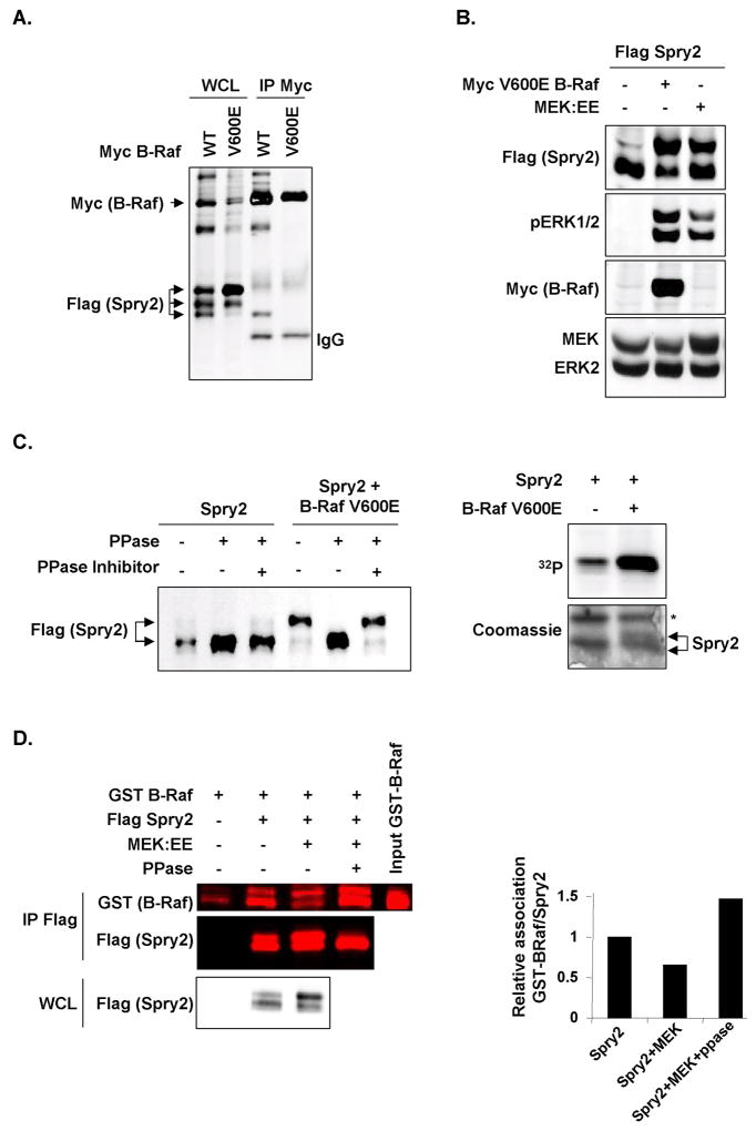
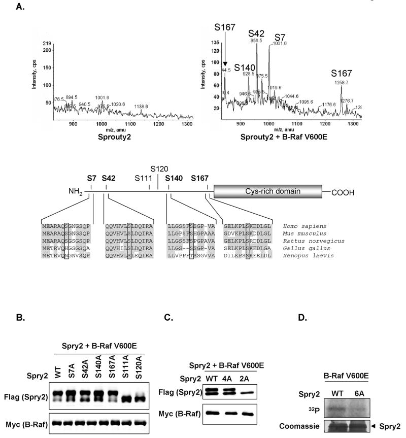
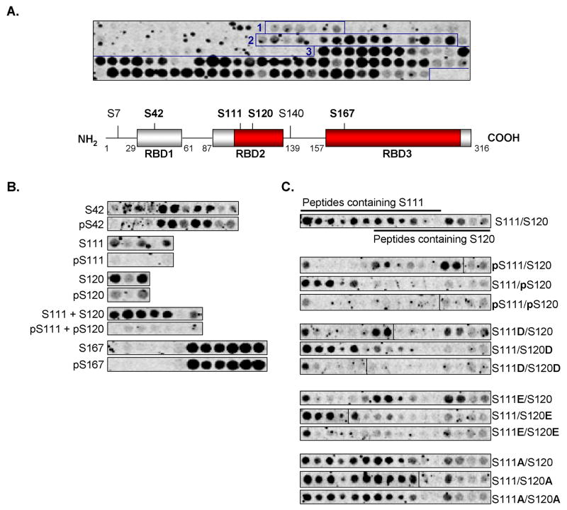
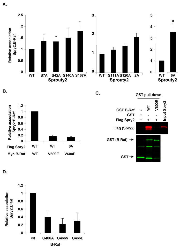
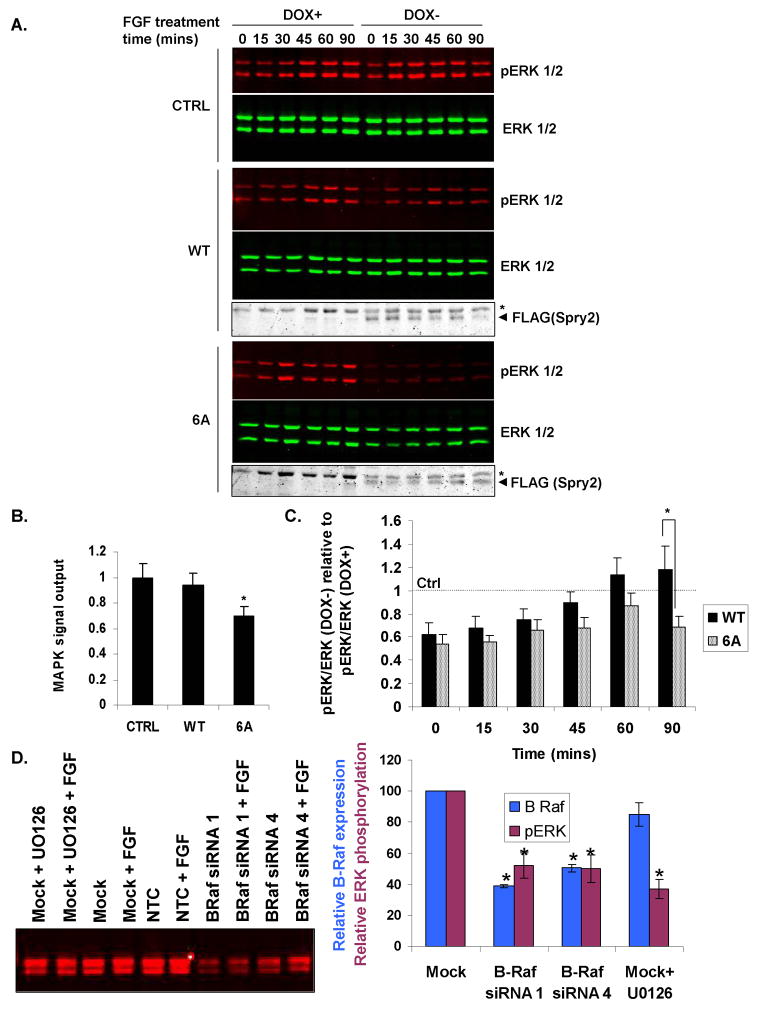
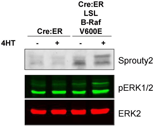
Sprouty2 is a feedback regulator that controls the Ras/Raf/MEK/extracellular signal-regulated kinase mitogen-activated protein kinase (MAPK) pathway at multiple levels, one way being through direct interaction with Raf kinases. Consistent with a role as a tumor suppressor, Sprouty2 expression is often down-regulated in human cancers. However, Sprouty2 is up-regulated in some cancers, suggesting the existence of posttranscriptional mechanisms that permit evasion of Sprouty2-mediated antitumorigenic properties. We report that MAPK activation induces Sprouty2 phosphorylation on six serine residues, which reduced Sprouty2 association with wild-type B-Raf. Mutation of these six serines to nonphosphorylatable alanines increased the ability of Sprouty2 to inhibit growth factor-induced MAPK activation. Oncogenic B-Raf mutants such as B-Raf V600E did not associate with Sprouty2, but this resistance to Sprouty2 binding was not due to phosphorylation. Instead, the active kinase conformation induced by oncogenic mutation prevents Sprouty2 binding. These results reveal a dual mechanism that affects the Sprouty2/B-Raf interaction: Sprouty phosphorylation and B-Raf conformation.


Figures

Figure 1
Figure 1
Sprouty2 phosphorylation regulates association with B-Raf. A, Cells were transfected with FLAG-Sprouty2 and Myc-B-Raf WT or V600E. WCL and Myc immunoprecipitates (IP Myc) were western blotted. B, Western blots of cells co-expresssing FLAG-Sprouty2 and Myc-B-Raf V600E or MEK1:EE. C, Left panel, FLAG-Sprouty2 transfected alone or with Myc-B-Raf V600E was immunoprecipitated and treated with CIP phosphatase (PPase) and phosphatase inhibitor as indicated. Right panel, Metabolic [32P] labeling of cells transfected with FLAG-Sprouty2 alone or with Myc-B-Raf V600E as indicated. Immunoprecipitated Sprouty2 was analyzed by autoradiography (upper panel) and Coomassie staining (lower panel). Asterisk=non-specific protein. D, FLAG-Sprouty2 transfected alone or with MEK1:EE was immunoprecipitated, the sample co-expressed with MEK1:EE was divided in two and treated with or without phosphatase as indicated. Immunoprecipitated Sprouty2 was then incubated with GST-B-Raf catalytic domain and recovered by anti-FLAG immunoprecipitation. Anti-FLAG IP (IP FLAG), WCL and input GST-B-Raf were western blotted for GST and/or FLAG.
Figure 2
Figure 2
B-Raf induces Sprouty2 phosphorylation on multiple serine residues. A, LC-MS analysis of Sprouty2 tryptic peptides with precursor 79 scanning on a 4000 Q-Trap mass spectrometer. Extracted ion chromatograms for the phosphopeptides detected from immunoprecipitated Sprouty2 without (left) or with (right) co-expressed B-Raf V600E, identities of each phosphorylation site shown. Serine 167 phosphopeptide ion was detected as the M-2H (1256.7) and M-3H (844.5) ions whereas the other three phosphopeptides were detected as M-2H ions. Schematic representation showing Sprouty2 phosphorylation sites identified by mass spectrometry in bold. Sequence alignment compares Sprouty2 homology surrounding these sites (highlighted by boxes) in selected species. Conserved Sprouty2 cysteine-rich domain and previously-identified sites Ser111 and Ser120 are shown. B, and C, Wild-type or phosphorylation site mutant FLAG-Sprouty2 co-expressed with B-Raf V600E were western blotted for FLAG and Myc. D, Recombinant GST-FLAG-Sprouty2 wild-type or 6A were incubated with WCL expressing Myc-B-Raf V600E in the presence of γ–[32P]-ATP and analyzed for [32P] incorporation by autoradiography (upper panel) and Coomassie staining (lower panel).
Figure 3
Figure 3
Sprouty2 phosphorylation within Raf Binding Domain 2 inhibits B-Raf binding. A, Upper panel, binding of [35S]-Methionine labeled in vitro transcribed/translated (IVTT) HA-tagged B-Raf to a Sprouty2 peptide array, comprised of 23-mer peptides each shifted by 2 residues. Three Raf-binding domains (RBD 1-3) were detected (boxed peptide spots). Lower panel, schematic representation of mouse Sprouty2 protein indicating RBD 1-3 (grey boxes) positions. Higher affinity B-Raf binding highlighted in red. Sprouty2 phosphorylation sites in bold. B, [35S]-Methionine labeled HA-tagged B-Raf binding to arrayed Sprouty2 peptides with serine or phosphoserine residues as indicated. C, [35S]-Methionine labeled HA-tagged B-Raf binding to arrayed Sprouty2 peptides with aspartate, glutamate or alanine substitutions at Ser111 and/or Ser120 as indicated.
Figure 4
Figure 4
Effect of mutating Sprouty2 phosphorylation sites on binding to wild-type or cancer associated B-Raf mutants. A, FLAG-Sprouty2 wild-type or phosphorylation site mutants were transfected with Myc-B-Raf V600E as indicated. Myc immunoprecipitates and WCL were western blotted for FLAG or Myc. Sprouty2 association with B-Raf expressed relative to total Sprouty2. Mean values ± standard error of the mean (SEM), normalized to Sprouty2 WT binding to B-Raf, are shown. For left panel n=7, middle panel n=3, right panel n=3. *=significant difference (P<0.05) from wild-type Sprouty2 binding, determined by Student's t test. B, FLAG-Sprouty2 wild-type or phosphorylation site mutants were transfected with Myc wild-type B-Raf or Myc-B-Raf V600E as indicated. Myc immunoprecipitates and WCL were western blotted for FLAG or Myc. Mean values ± SEM (n=12), normalized to binding of Sprouty2 WT to B-Raf WT, are shown. C, GST, GST-B-Raf wild-type or GST-B-Raf V600E were incubated with recombinant FLAG-Sprouty2, recovered on glutathione Sepharose beads (GST pull-down) and analyzed for B-Raf and associated Sprouty2 by western blotting for GST and FLAG. Input FLAG-Sprouty2 was loaded as a positive control. D, FLAG-Sprouty2 was transiently transfected with cancer-associated Myc-B-Raf G466 P-loop mutants as indicated. Myc immunoprecipitates and WCL were western blotted for FLAG or Myc. Mean values ± SEM (n=4), normalized to Sprouty2 binding to WT B-Raf.
Figure 5
Figure 5
Inhibition of FGF-stimulated ERK activation by Sprouty2 phosphorylation site mutant. A, NIH3T3 TETOFF FLAG-Sprouty2 control (CTRL), wild-type (WT; clone 2 shown) or 6 alanine (6A; clone 3 shown) cell lines were cultured with or without DOX, stimulated with FGF for times indicated and western blotted for pERK, ERK and FLAG-Sprouty2. Arrowhead=FLAG-Sprouty2. B, FGF-induced MAPK signal output following Sprouty2 induction relative to uninduced condition was determined from areas under the curve (AUC) of time-course experiments. pERK/ERK for DOX- (Sprouty2 induced) conditions expressed relative to pERK/ERK for DOX+ (Sprouty uninduced) conditions were plotted for each timepoint and AUC calculated using the Trapezoidal Rule. Mean values ± SEM, normalized to control clone, from four (control) or seven (WT and 6A) repetitions are shown. *=significant difference (P<0.05) from wild-type determined by Student's t test. C, Ratios of pERK/ERK for DOX- (Sprouty2 induced) conditions expressed relative to pERK/ERK for DOX+ (uninduced) for each timepoint for wild-type or 6A Sprouty2. Mean values ± SEM (n=7), normalized to control clone. *=significant difference (P<0.05) between WT and 6A determined by Student's t test. D, NIH3T3 cells were transfected were mock transfected or transfected with non-targeting control (NTC) or B-Raf siRNAs as indicated. After 48 hours, cells were treated with FGF as above for 45 minutes, either with or without 10 μM U0126. B-Raf expression levels were determined by quantitative Western blotting (left panel), and the relative expression of B-Raf (right, dark blue) and ratio of pERK/ERK (right, burgundy) determined for these conditions. Mean values ± SEM (n=6), normalized to mock transfectant. *=significant difference (P<0.01) from mock transfectant determined by Student's t test.
Figure 6
Figure 6
Mobility shift of endogenous Sprouty2 induced by endogenous V600E B-Raf. Mouse embryo fibroblasts expressing conditionally active Cre:ER alone or in combination with a heterozygous knock-in V600E B-Raf mutation preceded by a Lox-Stop-Lox (LSL) cassette were either left untreated or treated with 50 nM 4-hydroxytamoxifen (4HT) for 96 hours to induce Cre-mediated recombination as indicated. Sprouty2, pERK1/2 and ERK2 levels were determined by quantitative Western blotting

Similar articles

Cited by

References

    1. Hacohen N, Kramer S, Sutherland D, Hiromi Y, Krasnow MA. sprouty encodes a novel antagonist of FGF signaling that patterns apical branching of the Drosophila airways. Cell. 1998;92:253–63. - PubMed
    1. de Maximy AA, Nakatake Y, Moncada S, Itoh N, Thiery JP, Bellusci S. Cloning and expression pattern of a mouse homologue of drosophila sprouty in the mouse embryo. Mech Dev. 1999;81:213–6. - PubMed
    1. Tefft JD, Lee M, Smith S, et al. Conserved function of mSpry-2, a murine homolog of Drosophila sprouty, which negatively modulates respiratory organogenesis. Curr Biol. 1999;9:219–22. - PubMed
    1. Minowada G, Jarvis LA, Chi CL, et al. Vertebrate Sprouty genes are induced by FGF signaling and can cause chondrodysplasia when overexpressed. Development. 1999;126:4465–75. - PubMed
    1. Kim HJ, Bar-Sagi D. Modulation of signalling by Sprouty: a developing story. Nat Rev Mol Cell Biol. 2004;5:441–50. - PubMed

Publication types

MeSH terms

Substances