. 2009 Oct 16;284(42):28746-53.
doi: 10.1074/jbc.M109.032904. Epub 2009 Aug 18.

MqsR, a crucial regulator for quorum sensing and biofilm formation, is a GCU-specific mRNA interferase in Escherichia coli

Affiliations

MqsR, a crucial regulator for quorum sensing and biofilm formation, is a GCU-specific mRNA interferase in Escherichia coli

Yoshihiro Yamaguchi et al. J Biol Chem. .

Abstract

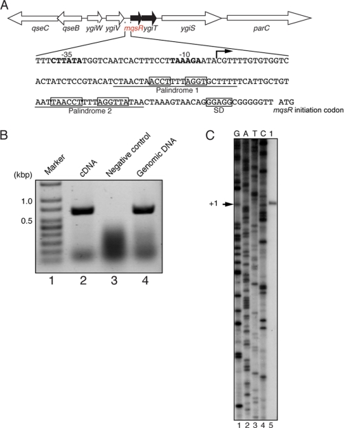
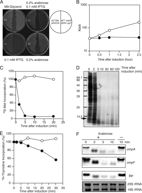
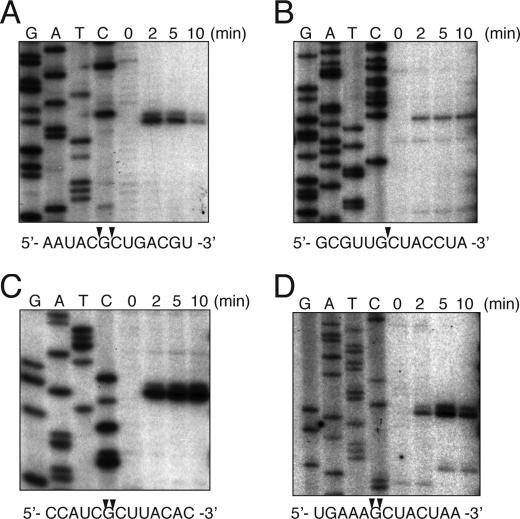
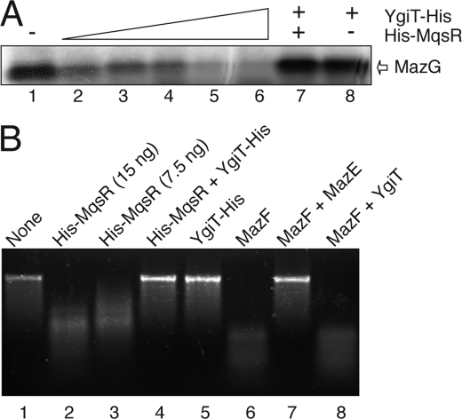
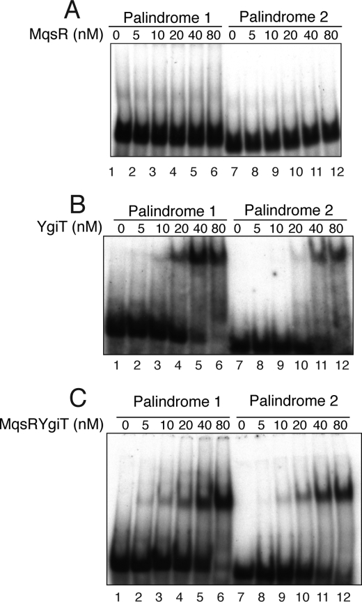
The mqsR gene has been shown to be positively regulated by the quorum-sensing autoinducer AI-2, which in turn activates a two-component system, the qseB-qseC operon. This operon plays an important role in biofilm formation in Escherichia coli. However, its cellular function has remained unknown. Here, we found that 1 base downstream of mqsR there is a gene, ygiT, that is co-transcribed with mqsR. Induction of mqsR caused cell growth arrest, whereas ygiT co-induction recovered cell growth. We demonstrate that MqsR (98 amino acid residues), which has no homology to the well characterized mRNA interferase MazF, is a potent inhibitor of protein synthesis that functions by degrading cellular mRNAs. In vivo and in vitro primer extension experiments showed that MqsR is an mRNA interferase specifically cleaving mRNAs at GCU. The mRNA interferase activity of purified MqsR was inhibited by purified YgiT (131 residues). MqsR forms a stable 2:1 complex with YgiT, and the complex likely functions as a repressor for the mqsR-ygiT operon by specifically binding to two different palindromic sequences present in the 5'-untranslated region of this operon.


Figures

FIGURE 1.
FIGURE 1.
Gene map of the mqsR-ygiT operon on the E. coli chromosome. A, arrows indicate the direction and size of the following genes: qseC, qseB, ygiW, ygiV, mqsR, ygiT, ygiS, and parC. The mqsR-ygiT promoter sequence is also shown, and the palindromic sequences (1 and 2) are boxed. The bent arrow represents the transcription initiation site of the mqsR-ygiT operon. The −10 and −35 regions of the mqsR-ygiT promoter are shown in boldface, and the Shine-Dalgarno (SD) sequence (GGAGG) is boxed. Underlined DNA sequences were used in electrophoretic mobility shift assay as shown in Fig. 5. B, shown are the results of reverse transcription-PCR analysis of the mqsR-ygiT operon. cDNA was synthesized with reverse transcriptase using total RNA from the E. coli BL21 strain grown at 37 °C to A600 = 0.8. Using the cDNA product as template, PCR was carried out with primers RT-Fw and RT-Rv. Lane 1, 100-bp DNA ladder (GenScript); lanes 2 and 4, cDNA and genomic DNA used as template for PCR, respectively; lane 3, PCR products without using reverse transcriptase. C, shown is the transcription start site of mqsR-ygiT. Primer extension analysis was carried out using the same RNA described for B and primer PX-RT. G, A, T, and C (lanes 1–4) comprise the sequence ladders using pCR®2.1-Topo®-mqsR-ygiT and the same primer. The transcription start site is indicated (+1).
FIGURE 2.
FIGURE 2.
Effect of MqsR induction on protein and DNA synthesis and mRNA stability. A, E. coli BL21 transformed with pET-mqsR and pBAD-ygiT and streaked on M9 (glycerol, CAA) plates with 0.1 mm isopropyl 1-thio-β-d-galactopyranoside (IPTG), with 0.2% arabinose, with 0.1 mm isopropyl 1-thio-β-d-galactopyranoside plus 0.2% arabinose, or without either inducer. The plates were incubated at 37 °C for 18 h. B, growth curves of E. coli BL21 cells harboring pBAD-mqsR. The cells were cultured in M9-glycerol liquid medium at 37 °C in the presence (●) or absence (○) of 0.2% arabinose. C, effect of MqsR on [35S]methionine incorporation in vivo. At the different time intervals indicated, 0.4 ml of the culture was put into a test tube containing 30 μCi of [35S]methionine, and the mixture was incubated for 30 s at 37 °C. After the incubation, 50 μl of the reaction mixture was applied to a filter paper disk (Whatman No. 3MM, 2.3-cm diameter). The filter paper disks were treated with 10% trichloroacetic acid solution as described previously (32). The radioactivity on the filter was determined with a liquid scintillation counter. D, SDS-PAGE analysis of the products in C. The reaction mixture (400 μl) at the time points indicated was put into a chilled test tube containing 100 μg/ml nonradioactive methionine, and cells were collected by centrifugation. The pellets were dissolved in 40 μl of SDS-PAGE loading buffer. The samples were incubated in a boiling water bath for 10 min. After removal of insoluble materials by centrifugation, the supernatant fraction (12.5 μl) was applied to 15% SDS-polyacrylamide gel. E, effect of MqsR on [3H]thymidine incorporation in vivo. E. coli BL21 cells harboring pBAD-mqsR were grown at 37 °C. When the A600 value of the culture reached 0.3, MqsR was induced with arabinose (0.2%). At the different time intervals indicated, 0.4 ml of the culture was put into a test tube and incubated with 10 μCi of [3H]thymidine plus 30 μg of nonradioactive thymidine. The mixture was then incubated for 30 s at 37 °C. After the incubation, the incorporated radioactivity into the cells were determined as described previously (32). F, effect of MqsR on cellular mRNA stability. Total RNA was extracted from E. coli BL21 cells harboring pBAD-mqsR at various time points as indicated after the addition of arabinose (0.2%) and subjected to Northern blotting with labeled ompA, ompF, and lpp as probes. Before transferring RNA onto a membrane, the gel was stained with ethidium bromide to detect 23 S and 16 S rRNAs.
FIGURE 3.
FIGURE 3.
Primer extension analysis of MqsR cleavage sites in ompF mRNA in vivo. Total RNA was prepared from E. coli BL21 cells harboring pBAD-mqsR at the indicated time points before and after the induction of MqsR. The sequence ladders were obtained with pCR®2.1-Topo®-ompF as template (32). The sequences around the cleavage sites are indicated below the panels, and the cleavage sites are indicated by arrowheads.
FIGURE 4.
FIGURE 4.
mRNA interferase activity of MqsR in vitro. A, effect of His-MqsR on protein synthesis in a cell-free system. MazG protein synthesis was carried out using an E. coli T7 S30 extract system for circular DNA with pET11a-mazG. Lane 1, without His-MqsR; lanes 2–6, 5, 10, 20, 40, and 80 nm His-MqsR, respectively; lane 7, 80 nm His-MqsR plus 40 nm YgiT-His; lane 8, 40 nm YgiT-His. B, mRNA interferase activity of purified His-MqsR in vitro. MS2 phage RNA (0.8 μg) was incubated with His-MqsR at 37 °C for 10 min in 10 mm Tris-HCl (pH 8.0) containing 1 mm dithiothreitol. The products were separated on a 1.2% agarose gel. The gel was stained with ethidium bromide.
FIGURE 5.
FIGURE 5.
Binding of MqsR, MqsR-YgiT, and YgiT to the palindromic sequences in the mqsR-ygiT 5′-UTR. Electrophoretic mobility shift assay was carried out with 5′-end-labeled palindrome 1 (lanes 1–6) and 2 (lanes 7–12) DNA fragments (see Fig. 1A), which were incubated with different concentrations of proteins as described under “Experimental Procedures.” Lanes 1–6 and 7–12 represent 0, 5, 10, 20, 40, and 80 nm His-MqsR (A), YgiT-His (B), and His-MqsR-YgiT (C), respectively.

References

    1. Balestrino D., Haagensen J. A., Rich C., Forestier C. (2005) J. Bacteriol. 187, 2870–2880 - PMC - PubMed
    1. Davies D. G., Parsek M. R., Pearson J. P., Iglewski B. H., Costerton J. W., Greenberg E. P. (1998) Science 280, 295–298 - PubMed
    1. Hammer B. K., Bassler B. L. (2003) Mol. Microbiol. 50, 101–104 - PubMed
    1. McNab R., Ford S. K., El-Sabaeny A., Barbieri B., Cook G. S., Lamont R. J. (2003) J. Bacteriol. 185, 274–284 - PMC - PubMed
    1. Ren D., Bedzyk L. A., Thomas S. M., Ye R. W., Wood T. K. (2004) Appl. Microbiol. Biotechnol. 64, 515–524 - PubMed

Publication types

LinkOut - more resources