. 2009 Nov;331(2):680-9.
doi: 10.1124/jpet.109.153601. Epub 2009 Aug 18.

Thioflavin S (NSC71948) interferes with Bcl-2-associated athanogene (BAG-1)-mediated protein-protein interactions

Affiliations

Thioflavin S (NSC71948) interferes with Bcl-2-associated athanogene (BAG-1)-mediated protein-protein interactions

Adam Sharp et al. J Pharmacol Exp Ther. 2009 Nov.

Abstract

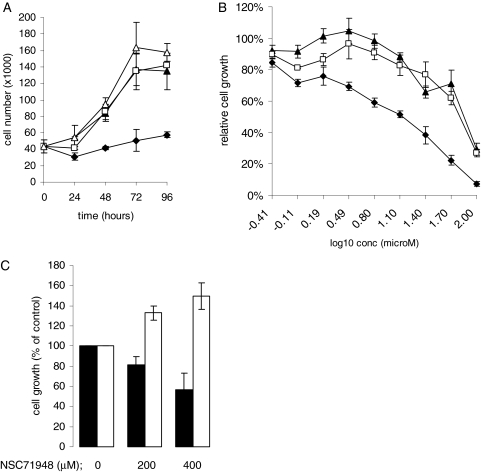
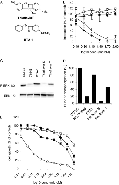
The C-terminal BAG domain is thought to play a key role in BAG-1-induced survival and proliferation by mediating protein-protein interactions, for example, with heat shock proteins HSC70 and HSP70, and with RAF-1 kinase. Here, we have identified thioflavin S (NSC71948) as a potential small-molecule chemical inhibitor of these interactions. NSC71948 inhibited the interaction of BAG-1 and HSC70 in vitro and decreased BAG-1:HSC70 and BAG-1:HSP70 binding in intact cells. NSC71948 also reduced binding between BAG-1 and RAF-1, but had no effect on the interaction between two unrelated proteins, BIM and MCL-1. NSC71948 functionally reversed the ability of BAG-1 to promote vitamin D3 receptor-mediated transactivation, an activity of BAG-1 that depends on HSC70/HSP70 binding, and reduced phosphorylation of p44/42 mitogen-activate protein kinase. NSC71948 can be used to stain amyloid fibrils; however, structurally related compounds, thioflavin T and BTA-1, had no effect on BAG-1:HSC70 binding, suggesting that structural features important for amyloid fibril binding and inhibition of BAG-1:HSC70 binding may be separable. We demonstrated that NSC71948 inhibited the growth of BAG-1 expressing human ZR-75-1 breast cancer cells and wild-type, but not BAG-1-deficient, mouse embryo fibroblasts. Taken together, these data suggest that NSC71948 may be a useful molecule to investigate the functional significance of BAG-1 C-terminal protein interactions. However, it is important to recognize that NSC71948 may exert additional "off-target" effects. Inhibition of BAG-1 function may be an attractive strategy to inhibit the growth of BAG-1-overexpressing cancers, and further screens of additional compound collections may be warranted.


Figures

Fig. 1.
Fig. 1.
Screening for inhibitors of the BAG-1:HSC70 interaction. A, human BAG-1 isoforms. The structures of the three major human BAG-1 isoforms are shown, along with their size (amino acid residues). Translation of BAG-1L initiates at an upstream CUG codon, whereas BAG-1M and BAG-1S are AUG-derived. The position of the nuclear localization sequence (NLS), acidic repeats, ubiquitin-like domain (ULD), and BAG domain are shown. B, diagram to demonstrate the principle of the screening assay. Negative controls included GST in place of GST-BAG-1S and wells lacking HSC70. C, overview of screening strategy. A total of 3156 compounds were screened in two sets of primary assays. Secondary assays to determine IC50 values resulted in identification of 16 confirmed hits. IC50 values for inhibition of BAG-1:HSC70 binding are shown (mean values derived from two independent experiments each performed in duplicate ± S.D.). *, compound selected for further study. The structures of the compounds listed are available from the National Cancer Institute Discovery Services (NCI-DTP) website (http://dtp.nci.nih.gov/webdata.html).
Fig. 2.
Fig. 2.
Identification of NSC71948 as an inhibitor of the in vitro BAG-1:HSC70 interaction. A, structure of NSC71948 (thioflavin S). B, inhibition of the in vitro BAG-1:HSC70 interaction by NSC71948. The experiment shows the mean of duplicate determinations (± S.D.) of BAG-1:HSC70 interaction in the presence of the indicated concentrations of NSC71948 (refilled stock). Experiment is representative of two independent experiments. C, luciferase counterscreen. The activity of recombinant luciferase was measured in the presence of NSC71948 or NSC119913 (both at 30 μM). The activity of luciferase in the absence of added compound was set at 100%. Data shown are mean (± S.D.) luciferase activity derived from two separate experiments, each performed in duplicate. D, BSA counterscreen. The ability of NSC71948 (30 μM) or NSC73413 (15 μM) to inhibit in vitro BAG-1:HSC70 binding was measured in the presence or absence of excess (0.1 mg/ml) BSA. Data shown are remaining activity (mean ± S.D.) in the presence of BSA and are derived from two separate experiments, each performed in duplicate.
Fig. 3.
Fig. 3.
Effect of NSC71948 on BAG-1:HSC70/HSP70 binding in intact MCF7 cells. A, MCF7-BAG-1S cells were incubated with 100 μM NSC71948, or left untreated as a control. Immunoprecipitations were performed by use of BAG-1-specific antibody TB3 from cells cultured in the presence or absence of NSC71948, or with control preimmune sera (PI). BAG-1S, HSC70, and HSP70 were analyzed by immunoblotting. B, the amounts of BAG-1-bound HSC70 and HSP70 were quantified by digital imaging. The graph shows the interaction of BAG-1 and HSC70 or HSP70 in the presence NSC71948. The level of binding in the absence of NSC71948 was used to set the 100% value. Data are mean (± S.D.) binding derived from two independent experiments each performed in duplicate. *, p <0.05 compared with untreated cells (Student's t test). C, MCF7 cells were incubated with 100 μM NSC71948, or left untreated as a control, and the binding of MCL-1 and BIM determined by coimmunoprecipitation/immunoblotting. The amount of MCL-1-bound BIM was determined by digital imaging and was set at 100% in control cells. The graph shows mean (± S.D.) MCL-1:BIM binding in the presence or absence of NSC71948. The data are derived from two independent experiments, each performed in duplicate.
Fig. 4.
Fig. 4.
Effect of NSC71948 on regulation of VDR by BAG-1L. A, H376 cells were transfected with a vitamin D3-responsive luciferase reporter construct and the indicated BAG-1L expression constructs, or pcDNA3 as a control. Cells were also transfected with a constant amount of renilla luciferase to control for transfection efficiency. After 2 days, cells were treated with NSC71948 as indicated, and cells were stimulated with vitamin D3 (100 nM) (■) or left untreated as a control (□). Firefly and renilla luciferase activity were determined after 24 h. The figure shows normalized luciferase activity relative to control, unstimulated, pCDNA3-transfected cells (set to 1.0). Data shown are the mean (± S.D.) of triplicate transfections and are representative of four individual experiments. B, immunoblot analysis of BAG-1L expression in H376 cells transfected with the BAG-1L expression plasmid, or pcDNA3 as a control. Cells were treated with NSC71948 or NSC119913 (50 μM) as indicated.
Fig. 5.
Fig. 5.
Effect of NSC71948 on ERK1/2 phosphorylation and BAG-1:RAF-1 binding. A, MCF7 cells were treated for 2 h with the indicated concentrations of NSC71948 or left untreated as a control. Expression of total and phosphorylated ERK1/2 was analyzed by immunoblotting. The data are representative of two experiments performed in duplicate. B, inhibition of ERK1/2 phosphorylation by NSC71948 was quantified by digital imaging. The data shown are mean (± S.D.) ERK1/2 phosphorylation derived from two independent experiments each performed in duplicate. C, MCF7 cells were treated with DMSO, NSC71948, or NSC119913 (both at 50 μM) for 16 h and immunoprecipitations performed by use of a preimmune (PI) or BAG-1-specific antisera (TB3). The expression of RAF-1 and BAG-1 in whole cell lysates (left) and immunoprecipitates (right) was detected by immunoblotting. The data shown are representative of four experiments.
Fig. 6.
Fig. 6.
Growth inhibition by NSC71948. A, ZR-75-1 cells were plated at a density of 20,000 cells/well of a 24-well plate and allowed to recover for 24 h. Cell number was determined (time, 0 h) and the remaining wells were treated with 50 μM NSC71948 (♦), 50 μM NSC119913 (▴), DMSO (□), or left untreated as control (▵). Cell numbers were determined for up to 4 days. Results shown are mean of duplicate determinations (± S.D.). B, ZR-75-1 cells were treated with the indicated concentrations of NSC71948 (♦), NSC119913 (▴), or DMSO (□). After 6 days, cell number was determined by use of the CellTiter assay. Values obtained for untreated cells were set to 100%. Data are mean of triplicate determinations (± S.D.) and are representative of three independent experiments. C, wild-type (■) and BAG-1-deficient (□) MEFs were treated with 200 or 400 μM NSC71948, or were left untreated as a control. After 3 days, cell number was determined by use of the CellTiter assay. Values obtained for untreated cells were set to 100%. Data are mean of two experiments (± S.D.), each performed in triplicate.
Fig. 7.
Fig. 7.
Analysis of structurally related compounds. (A) Structures of thioflavin S/NSC71948, thioflavin T and BTA1. (B) Inhibition of the in vitro BAG-1:HSC70 interaction. The experiment shows the mean (± S.D.) of duplicate determinations of BAG-1:HSC70 binding in the presence of the indicated concentrations of NSC71948 (♦), thioflavin S (×), thioflavin T (♢), BTA-1 (■), or DMSO (□). Binding in the absence of any added compound was set to 100%. The data are representative of two independent experiments. C, MCF7 cells were treated for 2 h with NSC71948, thioflavin S, thioflavin T, or BTA-1 (each at 50 μM), or DMSO as control. Expression of total and phosphorylated ERK1/2 was analyzed by immunoblotting. Data shown are representative of two experiments, each performed in duplicate. D, quantitation of effects of compounds (50 μM) on ERK1/2 phosphorylation. E, ZR-75-11 cells were treated with the indicated concentrations of NSC71948 (♦), thioflavin T (♢), BTA1 (■), or DMSO (□) as a control. After 6 days, cell number was determined by use of the CellTiter assay. Values obtained for untreated cells were set to 100%. Data are mean of triplicate determinations (± S.D.).

References

    1. Alberti S, Demand J, Esser C, Emmerich N, Schild H, Hohfeld J. (2002) Ubiquitylation of BAG-1 suggests a novel regulatory mechanism during the sorting of chaperone substrates to the proteasome. J Biol Chem 277:45920–45927 - PubMed
    1. Arndt V, Rogon C, Höhfeld J. (2007) To be, or not to be—molecular chaperones in protein degradation. Cell Mol Life Sci 64:2525–2541 - PMC - PubMed
    1. Briknarová K, Takayama S, Brive L, Havert ML, Knee DA, Velasco J, Homma S, Cabezas E, Stuart J, Hoyt DW, et al. (2001) Structural analysis of BAG1 cochaperone and its interactions with Hsc70 heat shock protein. Nat Struct Biol 8:349–352 - PubMed
    1. Brimmell M, Burns JS, Munson P, McDonald L, O'Hare MJ, Lakhani SR, Packham G. (1999) High level expression of differentially localized BAG-1 isoforms in some oestrogen receptor-positive human breast cancers. Br J Cancer 81:1042–1051 - PMC - PubMed
    1. Clemo NK, Collard TJ, Southern SL, Edwards KD, Moorghen M, Packham G, Hague A, Paraskeva C, Williams AC. (2008) BAG-1 is up-regulated in colorectal tumour progression and promotes colorectal tumour cell survival through increased NF-kappaB activity. Carcinogenesis 29:849–857 - PubMed

Publication types

MeSH terms