. 2009 Dec;23(12):4231-43.
doi: 10.1096/fj.09-138412. Epub 2009 Aug 18.

Activated Abl kinase inhibits oncogenic transforming growth factor-beta signaling and tumorigenesis in mammary tumors

Affiliations

Activated Abl kinase inhibits oncogenic transforming growth factor-beta signaling and tumorigenesis in mammary tumors

Tressa M Allington et al. FASEB J. 2009 Dec.

Abstract

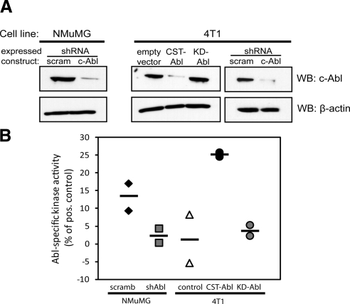
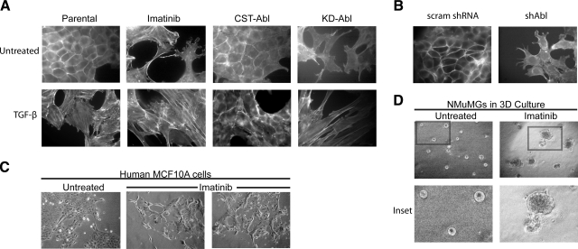
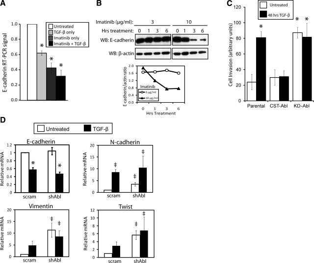
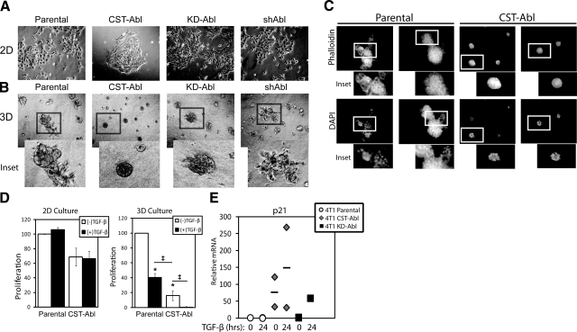
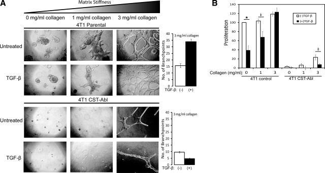
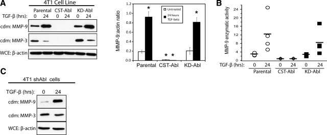
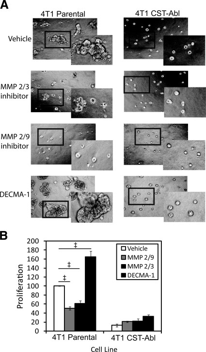
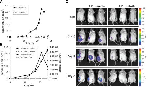
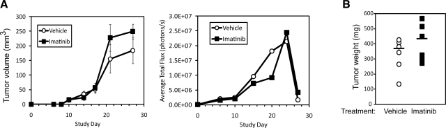
Transforming growth factor-beta (TGF-beta) is a ubiquitous cytokine with dual roles in tumor suppression and promotion, and these dichotomous functions have frustrated the development of therapies targeting oncogenic signaling by TGF-beta. In comparison, Abl is well established as an initiator of hematopoietic cancers; however, a clear role for Abl in regulating solid tumor development remains elusive. Here, we investigated the role of Abl in TGF-beta-mediated epithelial-mesenchymal transition (EMT) in normal and metastatic mammary epithelial cells (MECs). In doing so, we identified Abl as an essential regulator of MEC morphology and showed that Abl inactivation was sufficient to induce phenotypic and transcriptional EMT in normal MECs. Increasing Abl activity in metastatic MECs resulted in their complete morphological reversion, restored their cytostatic response to TGF-beta, and blocked their secretion of matrix metalloproteinases induced by TGF-beta. Constitutively active Abl expression blocked TGF-beta-responsive mammary tumor growth in mice, while Imatinib therapy afforded no clinical benefit in mice bearing mammary tumors. Collectively, this investigation establishes Abl as a potent mediator of MEC identity, and as a suppressor of oncogenic TGF-beta signaling during mammary tumorigenesis. Notably, our findings strongly caution against the use of pharmacological Abl antagonists in the treatment of developing and progressing mammary tumors.


Figures

Figure 1.
Figure 1.
Manipulation of Abl expression and activity in normal and malignant MECs. A) Immunoblotting for c-Abl expression in NMuMG and 4T1 cells engineered to express either a scrambled (scram) or an Abl-targeting shRNA (shAbl), or empty vector (pMSCV-hygromycin), CST-Abl, or KD-Abl, as indicated. Differences in protein loading were monitored by reprobing stripped membranes with anti-β-actin antibodies. B) ATP consumption assays were performed to compare Abl PTK activity in manipulated NMuMG and 4T1 cell lines. Horizontal lines show means of 2 independent experiments.
Figure 2.
Figure 2.
Abl inactivation morphologically mimics EMT induced by TGF-β in normal MECs. A) Direct actin immunofluorescence using FITC-conjugated phalloidin on Abl-manipulated NMuMG cells before and after their stimulation with TGF-β1 (5 ng/ml) or those treated with Imatinib (20 μg/ml) for 24 h as indicated. Images are representative of 3 independent experiments. B) FITC-conjugated phalloidin immunofluorescence on control (i.e., scrambled shRNA) or Abl-deficient NMuMG cells. Images are representative of 3 or 4 independent experiments. scram, scrambled shRNA; shAbl, Abl-targeting shRNA. C) Normal human MCF10A cells were cultured in the absence or presence of Imatinib (30 μg/ml) for 56 h, at which point bright-field images (×70) were collected. Data are representative of 2 independent experiments. D) NMuMG cells were cultured in compliant 3-D organotypic cultures in the absence or presence of Imatinib (20 μg/ml) for 5 d, at which point bright-field images (×70) were collected. Insets: magnified views of boxed regions. Data are representative of 3 independent experiments.
Figure 3.
Figure 3.
Abl inactivation transcriptionally mimics EMT and cell invasion induced by TGF-β in normal MECs. A) Quiescent NMuMG cells were treated with Imatinib (10 μg/ml), TGF-β1 (5 ng/ml), or both agents for 24 h as indicated. Afterward, relative levels of E-cadherin mRNA were detected by semiquantitative real-time PCR and normalized to corresponding GAPDH signals. Data are expressed as means ± se; n = 3. B) Quiescent NMuMG cells were incubated with increasing concentrations of Imatinib (3–10 μg/ml) for 0–6 h as indicated. Afterward, altered E-cadherin expression was monitored by immunoblotting with anti-E-cadherin antibodies, followed by immunoblotting for β-actin to monitor differences in protein loading. Accompanying graph depicts ratio of E-cadherin/β-actin signals. Data are from a representative experiment that was performed 3 times with similar results. C) Parental, CST-Abl-, or KD-Abl-expressing NMuMG cells were induced to invade synthetic basement membranes by a 4% serum stimulus, or by 4% serum plus TGF-β1 (5 ng/ml) for 48 h. Data are means ± se; n = 2. D) Quiescent NMuMG cells expressing either a scrambled (scram) or Abl-targeting shRNA (shAbl) were stimulated with TGF-β1 (5 ng/ml) for 48 h. Afterward, the relative mRNA expression levels of E- and N-cadherins, vimentin, and Twist were determined by semiquantitative real-time PCR. Data are means ± se; n = 3. *P < 0.01, P < 0.05 vs. corresponding control; Student’s t test).
Figure 4.
Figure 4.
Abl activation promotes normal MEC morphology and TGF-β-mediated cytostasis in metastatic MECs grown in compliant microenvironments. A, B) Bright-field images (×70) of parental, CST-Abl-, KD-Abl, or shAbl-expressing 4T1 cells grown in 2-D (A) or compliant 3-D organotypic (B) culture systems as indicated. Insets: magnified views of boxed regions. Images are representative of 3 independent experiments. C) Parental or CST-Abl-expressing 4T1 cells were grown in compliant 3-D organotypic cultures for 7 d as indicated. Afterward, resulting acinar structures were stained with FITC-phalloidin and DAPI. Insets: magnified views of boxed regions. Images are representative of 2 independent experiments. D) Parental or CST-Abl-expressing 4T1 cells were stimulated with TGF-β1 (5 ng/ml) for 48 h in 2-D (left panel) or compliant 3-D organotypic (right panel) cultures as indicated. Differences in proliferation were determined by employment of MTS proliferation assays. Data are means ± se; n = 2–5. *P < 0.01, P < 0.05 vs. untreated parental 4T1 cells; Student’s t test. E) 4T1 cells expressing empty vector, CST-Abl, or KD-Abl were stimulated with TGF-β1 (5 ng/ml) for 24 h. Afterward, relative mRNA expression of p21Cip1 transcripts was determined by semiquantitative real-time PCR. Data are combined from 2 independent experiments.
Figure 5.
Figure 5.
Abl activation abrogates rigidity-induced proliferation and maintains TGF-β cytostatic response in metastatic MECs. Parental and CST-Abl-expressing 4T1 cells were grown in the absence or presence of TGF-β1 (5 ng/ml) for 7 d in compliant or rigid (i.e., 1–3 mg/ml type I collagen) 3-D organotypic cultures as indicated. A) Left panels: differences in cell morphology and proliferation were monitored by bright-field microscopy (×70). Right panels: differences in organoid branching at 3 mg/ml collagen. B) Differences in organoid proliferation were monitored by MTS assays. Data are expressed as means ± se (n=3). *P < 0.01, P < 0.05 vs. untreated parental 4T1 cells; Student’s t test.
Figure 6.
Figure 6.
Abl activation alleviates basal and TGF-β-induced MMP expression and secretion. A) Quiescent parental, CST-Abl-, and KD-Abl-expressing 4T1 cells were stimulated with TGF-β1 (5 ng/ml) for 24 h, at which point the resulting conditioned medium (cdm) was collected and immunoblotted for MMPs 9 and 3 as indicated. Differences in cell numbers produced were monitored by immunoblotting for β-actin in corresponding whole-cell extracts (WCE). Graph depicts mean ± se ratios of MMP-9/β-actin signals; n = 5. *P < 0.01; Student’s t test. B) Conditioned medium was prepared as in A and was immunoprecipitated with anti-MMP-9 antibodies for use in fluorescent MMP-9 enzymatic assays. Symbols represent data obtained in 4 independent experiments; horizontal bars depict mean MMP-9 activity. C) Abl-deficient 4T1 cells (shAbl) were stimulated with TGF-β1 (5 ng/ml) for 24 h, at which point conditioned medium was collected and immunoblotted for MMPs 9 and 3 as indicated. Differences in cell numbers were monitored by immunoblotting corresponding whole-cell extracts with anti-β-actin antibodies. Images are from a representative experiment that was performed 2 times with identical results.
Figure 7.
Figure 7.
MMP and E-cadherin inhibitors alter the morphology and proliferation of metastatic MECs in 3-D organotypic cultures. Parental and CST-Abl-expressing 4T1 cells were cultured in the absence or presence of an MMP 2/3 inhibitor (30 μM), an MMP 2/9 inhibitor (500 nM), or a neutralizing E-cadherin antibody (DECMA-1; 0.5 mg/ml) for 5 d in compliant 3-D organotypic cultures as indicated. Differences in cell morphology and proliferation were monitored by bright field microscopy (×70; A), or by MTS proliferation assays (B). Insets: magnified views of boxed regions. Data are means ± se; n = 3. P < 0.05 vs. untreated parental 4T1 cells; Student’s t test).
Figure 8.
Figure 8.
Abl activation prevents 4T1 tumor growth in mice. A) Parental and CST-Abl-expressing 4T1 cells were injected orthotopically (103 cells/injection) into female Balb/C mice (12 animals/cell line), and 4T1 tumor growth was measured using digital calipers. B) Luciferase-expressing parental and CST-Abl-expressing 4T1 cells were engrafted onto the mammary fat pads of Balb/C mice (6 mice/cell line) as in A. Primary tumor growth was monitored using digital calipers (squares) or bioluminescent imaging (circles) measured as total flux of light emitted from imaged animals. C) Representative images of parental and CST-Abl-expressing 4T1 tumor-bearing mice imaged on d 0, 10, 17, and 21 postengraftment.
Figure 9.
Figure 9.
Imatinib administration fails to inhibit 4T1 tumor growth in mice. A) Luciferase-expressing 4T1 cells were injected orthotopically (103 cells/injection) into female Balb/C mice. Palpable tumors were readily detected in all mice at d 8 postinjection, at which point 6 mice received vehicle (i.e., sterile water) and 6 mice received Imatinib (50 mg/kg/d; oral administration). Primary tumor growth was monitored using digital calipers (left panel; means ± se) or bioluminescent imaging (right panel) measured as average total flux of light emitted from imaged animals. B) Primary 4T1 tumors were surgically removed at d 24 and 27 postengraftment and weighed. Bars indicate mean tumor weight for each group (6 mice/group).

References

    1. Benson J R. Role of TGF-β in breast carcinogenesis. Lancet Oncol. 2004;5:229–239. - PubMed
    1. Dumont N, Arteaga C L. TGF-β and breast cancer: Tumor promoting effects of TGF-β. Breast Cancer Res. 2000;2:125–132. - PMC - PubMed
    1. Pardali K, Moustakas A. Actions of TGF-β as tumor suppressor and pro-metastatic factor in human cancer. Biochim Biophys Acta. 2007;1775:21–62. - PubMed
    1. Wakefield L M, Piek E, Bottinger E P. TGF-β signaling in mammary gland development and tumorigenesis. J Mammary Gland Biol Neoplasia. 2001;6:67–82. - PubMed
    1. Wakefield L M, Roberts A B. TGF-β signaling: positive and negative effects on tumorigenesis. Curr Opin Genet Dev. 2002;12:22–29. - PubMed

Publication types