. 2009 Dec;23(12):4335-43.
doi: 10.1096/fj.09-137778. Epub 2009 Aug 18.

All of the human beta-type globin genes compete for LCR enhancer activity in embryonic erythroid cells of yeast artificial chromosome transgenic mice

Affiliations

All of the human beta-type globin genes compete for LCR enhancer activity in embryonic erythroid cells of yeast artificial chromosome transgenic mice

Eiichi Okamura et al. FASEB J. 2009 Dec.

Abstract

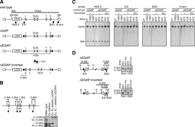
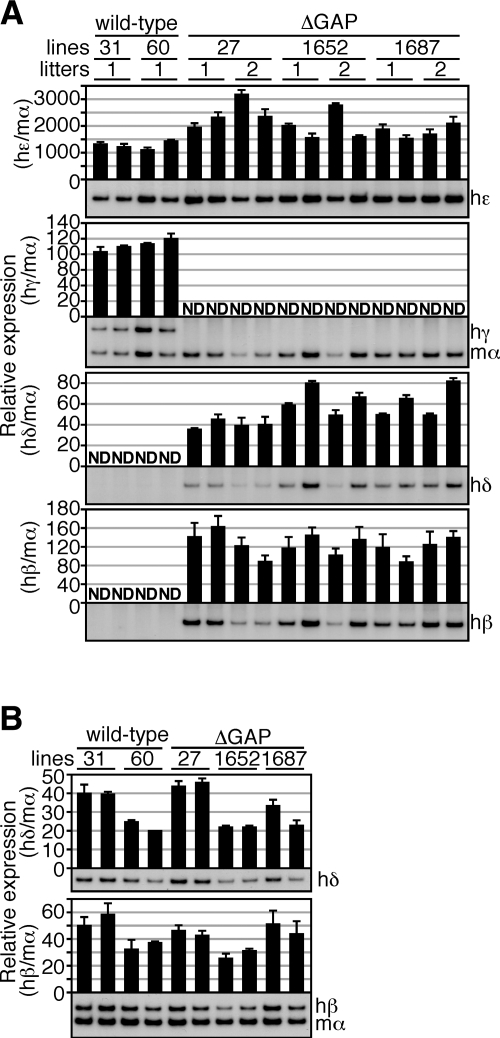
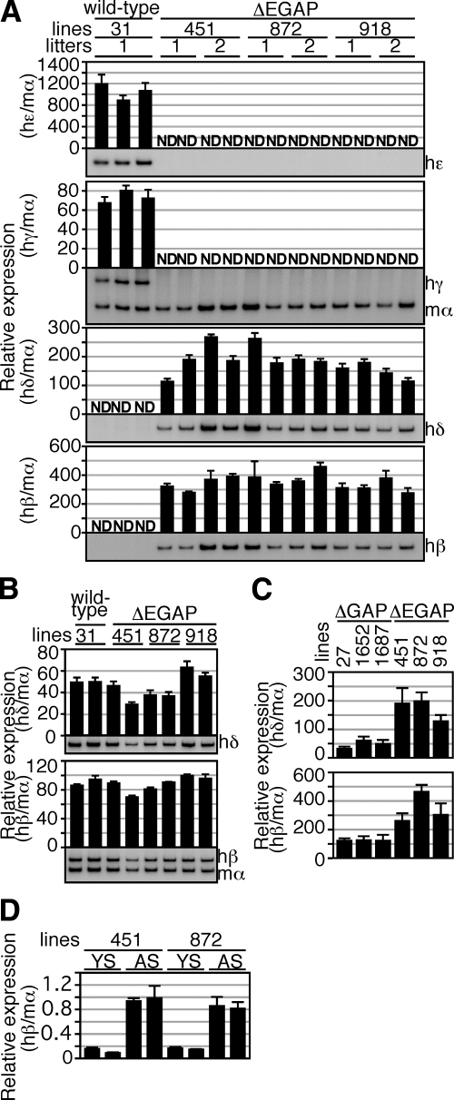
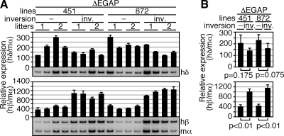
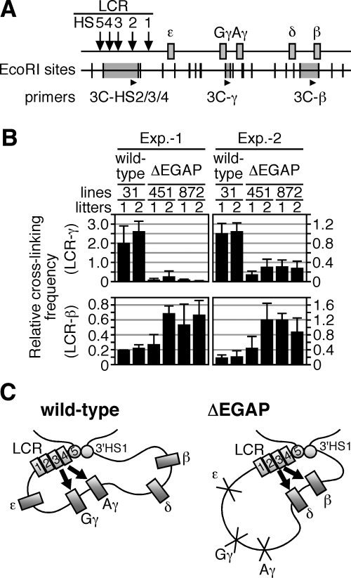
In primitive erythroid cells of human beta-globin locus transgenic mice (TgM), the locus control region (LCR)-proximal epsilon- and gamma-globin genes are transcribed, whereas the distal delta- and beta-globin genes are silent. It is generally accepted that the beta-globin gene is competitively suppressed by gamma-globin gene expression at this developmental stage. Previously, however, we observed that epsilon-globin gene expression was severely attenuated when its distance from the LCR was extended, implying that beta-globin gene might also be silenced because of its great distance from the LCR. Here, to clarify the beta-globin gene silencing mechanism, we established TgM lines carrying either gamma- or epsilon- plus gamma-globin promoter deletions, without significantly altering the distance between the beta-globin gene and the LCR. Precocious expression of delta- and beta-globin genes was observed in primitive erythroid cells of mutant, but not wild-type TgM, which was most evident when both the epsilon and gamma promoters were deleted. Thus, we clearly demonstrated that the repression of the delta- and beta-globin genes in primitive erythroid cells is dominated by competitive silencing by the epsilon- and gamma-globin gene promoters, and that epsilon- and the other beta-like globin genes might be activated by two distinct mechanisms by the LCR.


Figures

Figure 1.
Figure 1.
Experimental strategy. A) Schematic representation of the 150-kb wild-type and mutant human β-globin YACs. Positions of the β-like globin genes (gray rectangles) are shown relative to the LCR (open rectangle). SfiI restriction enzyme sites are located 5′ to HS5, between HS4 and HS3 of the LCR, and in the right arm of the YAC. Probes (solid rectangles) used for long-range fragment analysis shown in panel C and expected restriction enzyme fragments with their sizes are shown. In ΔGAP YAC, the Gγ (from −435 to +29 relative to transcriptional start site) and Aγ (from −435 to +29) promoters are missing (cross marks). In ΔEGAP YAC, the ε (from −438 to + 25), Gγ, and Aγ promoters are missing. In both mutant loci, loxP sites (arrowheads) were introduced in inverted orientation. The ΔEGAP-inverted locus was created by Cre-loxP recombination from the ΔEGAP locus in vivo. Distances between the LCR and β-globin gene in each locus are depicted. B) Introduction of the promoter mutations by homologous recombination in yeast. Partial restriction enzyme map of the wild-type human β-globin locus. X, XbaI; E, EcoRI. Probes and expected restriction enzyme fragments with their sizes are shown as solid and open rectangles, respectively. DNA from yeast clones bearing the wild-type or mutant human β-globin YACs was digested with XbaI and EcoRI and separated on agarose gels. After Southern blot transfer to nylon membrane, the DNA was hybridized to the mixed probes. Gγ-, Aγ-, and ε-globin gene promoters were sequentially deleted in this order. C) Long-range structural analysis of the human β-globin YAC in TgM. The whole β-globin locus is contained within 2 SfiI fragments (8 and 100 kb, shown in panel A). DNA from thymus cells of transgenic mice was digested with SfiI in agarose plugs, separated by pulsed-field gel electrophoresis, and Southern blots were hybridized separately to the probes. −, noninverted; inv., inverted. D) Cre-loxP-mediated in vivo inversion of the locus. Detailed structures around the loxP sites in the ΔEGAP and ΔEGAP-inverted transgenes are shown. Positions of the loxP sites (arrowheads), genes (labeled boxes), restriction enzyme sites (N, NcoI; P, PstI), and probes (solid rectangles) are shown proportionally. Expected restriction enzyme fragments with their sizes are shown. Tail DNAs from each mutant TgM line were digested with NcoI or PstI, separated on agarose gels, and Southern blots were hybridized to probes E3I or B3O.
Figure 2.
Figure 2.
Expression of the human β-like globin genes in ΔGAP TgM. A) Total RNA was prepared from the yolk sacs of >2 embryos (9.5 dpc) derived from the intercross of male TgM and female wild-type animals. Samples were collected from 2 independent litters of each mutant line. Expression of human ε (hε)-, γ (hγ)-, δ (hδ)-, and β (hβ)-globin compared to endogenous mouse α (mα)-globin genes was separately analyzed by semiquantitative RT-PCR. Signals for hε-globin at 18 cycles, hγ/mα-globin at 12 cycles, hδ-globin at 24 cycles, and hβ-globin at 18 cycles were quantified by PhosphoImager, and ratios of hε/mα, hγ/mα, hδ/mα, and hβ/mα were calculated (mα signal at 12 cycles was set at 100%). B) Total RNA was prepared from the spleens of 1- to 2-mo-old anemic mice. Samples were collected from 2 individuals from each line of TgM. Expression of hδ- and hβ-globin compared to endogenous mα-globin genes was separately analyzed by semiquantitative RT-PCR. Signals for hδ-globin at 16 cycles and hβ/mα-globin at 12 cycles were quantified by PhosphoImager, and ratios of hδ/mα and hβ/mα were calculated (mα signal at 12 cycles was set at 100%). Averages ± sd from ≥3 independent experiments were calculated and are graphically depicted. Representative results are shown at bottom of each panel.
Figure 3.
Figure 3.
Expression of the human β-like globin genes in ΔEGAP TgM. A) Total RNA was prepared from the yolk sacs of >2 embryos (9.5 dpc) from 2 independent litters of each mutant line. Expression of hε-, hγ-, hδ-, and hβ-globin compared to endogenous mα-globin genes was separately analyzed by semiquantitative RT-PCR, as described in Fig. 2A. B) Total RNA was prepared from the spleens of 1- to 2-mo-old anemic mice. Samples were collected from 2 individuals from each line of TgM. Expression of hδ- and hβ-globin compared to endogenous mα-globin genes was separately analyzed by semiquantitative RT-PCR, as described in Fig. 2B. C) Comparison of human β-like globin genes expression in the yolk sacs of ΔGAP and ΔEGAP TgM. RNA samples were analyzed by semiquantitative RT-PCR. Averages ± sd of ratios of hδ/mα (top) and hβ/mα (bottom) from 4 individuals of each mutant line were calculated and are graphically depicted. D) Comparison of hβ-globin gene expression in between the yolk sac (YS) and adult spleen (AS) of ΔEGAP TgM. RNA samples were analyzed by real-time PCR. Averages ± sd of the ratio of hβ/mα from ≥3 independent experiments were calculated and are graphically depicted.
Figure 4.
Figure 4.
Expression of the human β-like globin genes in ΔEGAP-inverted TgM. A) Total RNA was prepared from the yolk sacs of >2 embryos (9.5 dpc) from 2 independent litters of each mutant line. Expression of hδ- and hβ-globin compared to endogenous mα-globin genes was separately analyzed by semiquantitative RT-PCR as described in Fig. 2A. −, noninverted; inv., inverted. B) Comparison of human β-like globin gene expression between the yolk sacs of ΔEGAP and ΔEGAP-inverted TgM. RNA samples were analyzed by semiquantitative RT-PCR. Averages ± sd of the ratios of hδ/mα (top) and hβ/mα (bottom) from 4 individuals of each mutant line were calculated and are graphically depicted.
Figure 5.
Figure 5.
Spatial organization of the transgenic human β-globin locus. A) Strategy of the 3C assay in the human β-globin locus. Arrows and gray boxes represent the HS sites and the human β-like globin genes, respectively. EcoRI restriction enzyme sites (vertical lines) and position of the primers used for real-time qPCR (arrowheads) are shown. The 3C-HS2/3/4 primer was designed in the EcoRI fragment (gray highlight) containing HS2–4 of the LCR; 3C-γ and 3C-β primers in the regions around Gγ/Aγ-globin genes and β-globin gene, respectively. B) Comparison of the spatial organization between wild-type and ΔEGAP human β-globin loci in primitive erythroid cells. Chromatin from embryonic blood cells (10.5 dpc) was cross-linked with formaldehyde, digested with EcoRI, and religated. After reversal of cross-linking, purified DNA was subjected to real-time qPCR to determine cross-linking efficiencies between the LCR and γ- (top) or β-globin (bottom) gene regions. To control for digestion and religation efficiencies, as well as the amount of template DNA, between separately processed samples, values were normalized to those obtained with unrelated mouse Ercc3 gene primers . Blood samples were collected from 2 independent sets of animals (Exp. 1 and 2). Averages ± sd from ≥4 independent PCR reactions were calculated and are graphically depicted. C) Model of competitive silencing of the β-like globin genes at primitive stage of erythropoiesis. Throughout development, the β-like globin genes, together with the LCR and 3′HS1 (numbered rectangles and circles) are thought to form an active chromatin hub structure . In primitive erythroid cells, the spatial organization of individual human β-like globin genes (gray rectangles) relative to the LCR is different between wild-type and ΔEGAP loci. In wild-type TgM (left panel), active γ-globin genes are proximal to the LCR, while δ- and β- globin genes are looped out and silenced. In contrast, in ΔEGAP TgM (right panel), because the interaction with γ-globin gene promoters is disrupted, the LCR can now contact δ- and β-globin genes and potentiate transcription of these genes. Arrows indicate activation by LCR.

Similar articles

Cited by

References

    1. Stamatoyannopoulos G. Philadelphia/London: W. B. Saunders; The Molecular Basis of Blood Diseases. 1994
    1. Tuan D, Solomon W, Li Q, London I M. The “beta-like-globin” gene domain in human erythroid cells. Proc Natl Acad Sci U S A. 1985;82:6384–6388. - PMC - PubMed
    1. Grosveld F, van Assendelft G B, Greaves D R, Kollias G. Position-independent, high-level expression of the human beta-globin gene in transgenic mice. Cell. 1987;51:975–985. - PubMed
    1. Stamatoyannopoulos G. Control of globin gene expression during development and erythroid differentiation. Exp Hematol. 2005;33:259–271. - PMC - PubMed
    1. Raich N, Enver T, Nakamoto B, Josephson B, Papayannopoulou T, Stamatoyannopoulos G. Autonomous developmental control of human embryonic globin gene switching in transgenic mice. Science. 1990;250:1147–1149. - PubMed

Publication types