

The effect on cerebral tissue oxygenation index of changes in the concentrations of inspired oxygen and end-tidal carbon dioxide in healthy adult volunteers
- PMID: 19690266
- PMCID: PMC2742623
- DOI: 10.1213/ane.0b013e3181aedcdc
The effect on cerebral tissue oxygenation index of changes in the concentrations of inspired oxygen and end-tidal carbon dioxide in healthy adult volunteers
Abstract
Background: A variety of near-infrared spectroscopy devices can be used to make noninvasive measurements of cerebral tissue oxygen saturation (ScO2). The ScO2 measured by the NIRO 300 spectrometer (Hamamatsu Photonics, Japan) is called the cerebral tissue oxygenation index (TOI) and is an assessment of the balance between cerebral oxygen delivery and utilization. We designed this study to investigate the effect of systemic and intracranial physiological changes on TOI.
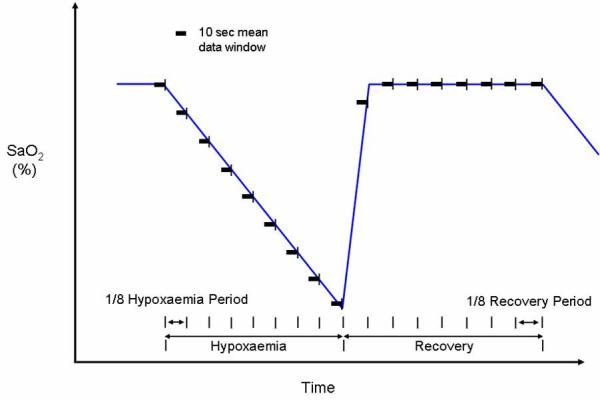
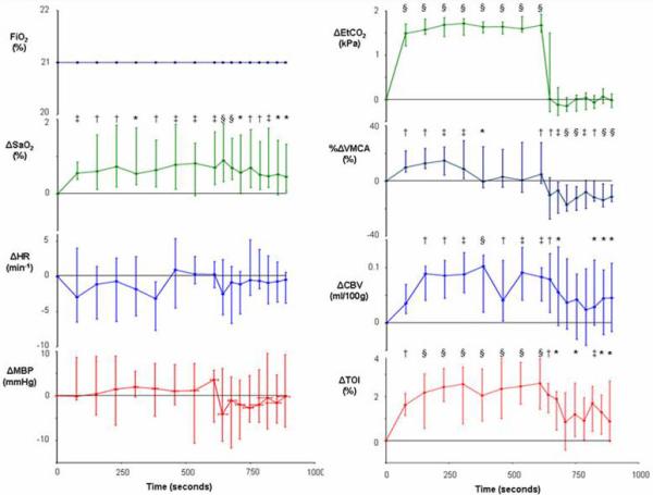
Methods: Fifteen healthy volunteers were studied during isocapneic hyperoxia and hypoxemia, and normoxic hypercapnea and hypocapnea. Absolute cerebral TOI and changes in oxy- and deoxyhemoglobin concentrations were measured using a NIRO 300 spectrometer. Changes in arterial oxygen saturation (SaO2), ETCO2, heart rate, mean arterial blood pressure (MBP), and middle cerebral artery blood flow velocity (Vmca) were also measured during these physiological challenges. Changes in cerebral blood volume (CBV) were subsequently calculated from changes in total cerebral hemoglobin concentration.
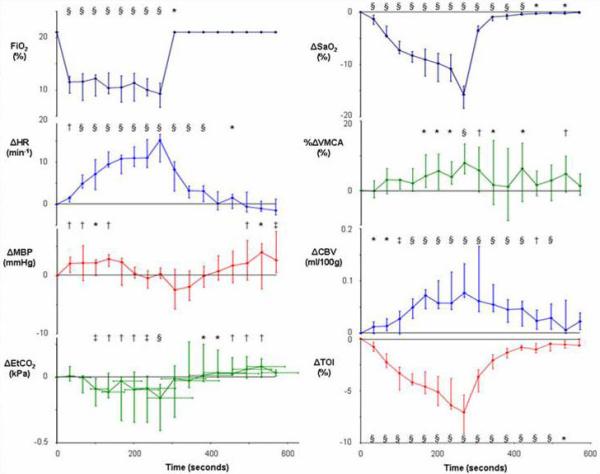
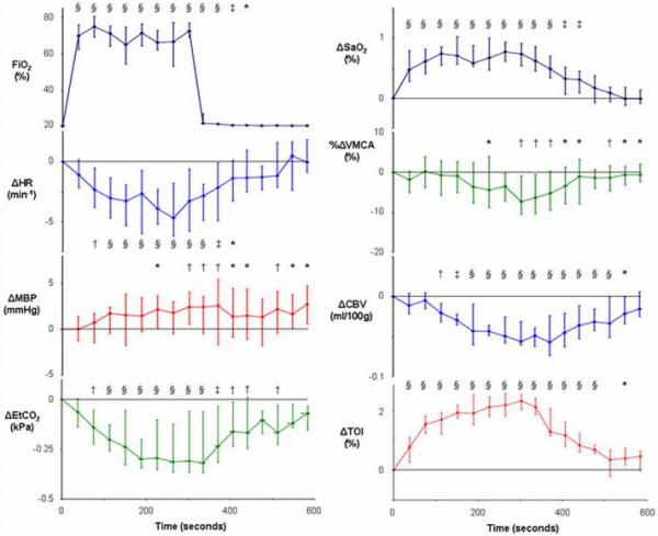
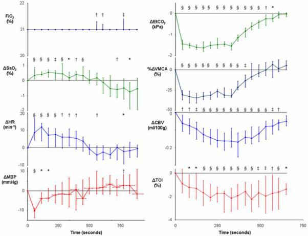
Results: Baseline TOI was 67.3% with an interquartile range (IQR) of 65.2%-71.9%. Hypoxemia was associated with a median decrease in TOI of 7.1% (IQR -9.1% to -5.4%) from baseline (P < 0.0001) and hyperoxia with a median increase of 2.3% (IQR 2.0%-2.5%) (P < 0.0001). Hypocapnea caused a reduction in TOI of 2.1% (IQR -3.3% to -1.3%) from baseline (P < 0.0001) and hypercapnea an increase of 2.6% (IQR 1.4%-3.7%) (P < 0.0001). Changes in SaO2 (P < 0.0001), ETCO2 (P < 0.0001), CBV (P = 0.0003), and MBP (P = 0.03) were significant variables affecting TOI. Changes in Vmca (P = 0.7) and heart rate (P = 0.2) were not significant factors.
Conclusion: TOI is an easy-to-monitor variable that provides real-time, multisite, and noninvasive assessment of the balance between cerebral oxygen delivery and utilization. However, TOI is a complex variable that is affected by SaO2 and ETCO2, and, to a lesser extent, by MBP and CBV. Clinicians need to be aware of the systemic and cerebral physiological changes that can affect TOI to interpret changes in this variable during clinical monitoring.
Figures
References
-
- Schell RM, Cole DJ. Cerebral monitoring: jugular venous oximetry. Anesth Analg. 2000;90:559–66. - PubMed
-
- Gupta AK, Hutchinson PJ, al-Rawi P, Gupta S, Swart M, Kirkpatrick PJ, Menon DK, Datta AK. Measuring brain tissue oxygenation compared with jugular venous oxygen saturation for monitoring cerebral oxygenation after traumatic brain injury. Anesth Analg. 1999;88:549–53. - PubMed
-
- Tisdall MM, Smith M. Multimodal monitoring in traumatic brain injury: current status and future directions. Br J Anaesth. 2007;99:61–7. - PubMed
-
- Okada E, Delpy DT. Near-infrared light propagation in an adult head model. II. Effect of superficial tissue thickness on the sensitivity of the near-infrared spectroscopy signal. Appl Opt. 2003;42:2915–22. - PubMed
-
- Delpy DT, Cope M, van der Zee P, Arridge S, Wray S, Wyatt J. Estimation of optical pathlength through tissue from direct time of flight measurement. Phys Med Biol. 1988;33:1433–42. - PubMed
Publication types
MeSH terms
Substances
Grants and funding
LinkOut - more resources
Full Text Sources
Miscellaneous
