. 2009 Oct 16;284(42):29205-17.
doi: 10.1074/jbc.M109.043752. Epub 2009 Aug 18.

Structure-function studies of claudin extracellular domains by cysteine-scanning mutagenesis

Affiliations

Structure-function studies of claudin extracellular domains by cysteine-scanning mutagenesis

Susanne Angelow et al. J Biol Chem. .

Abstract

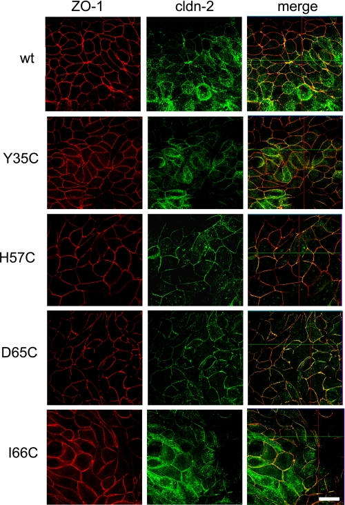
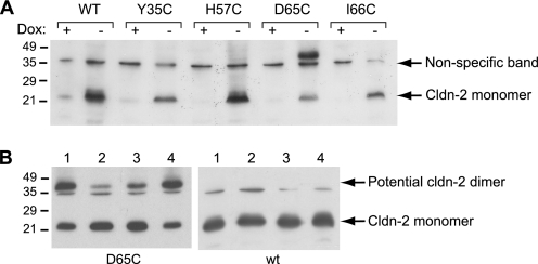
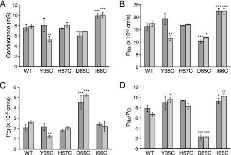
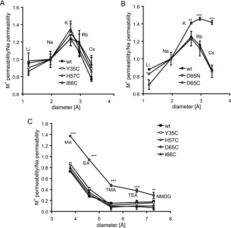
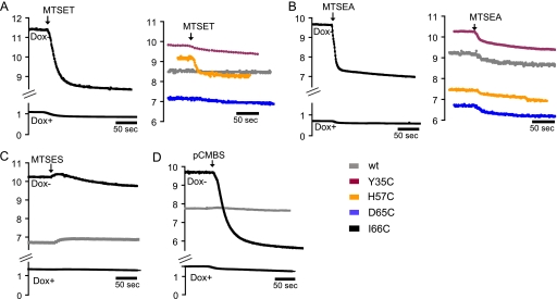
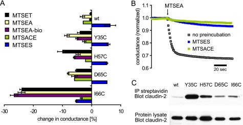
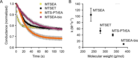
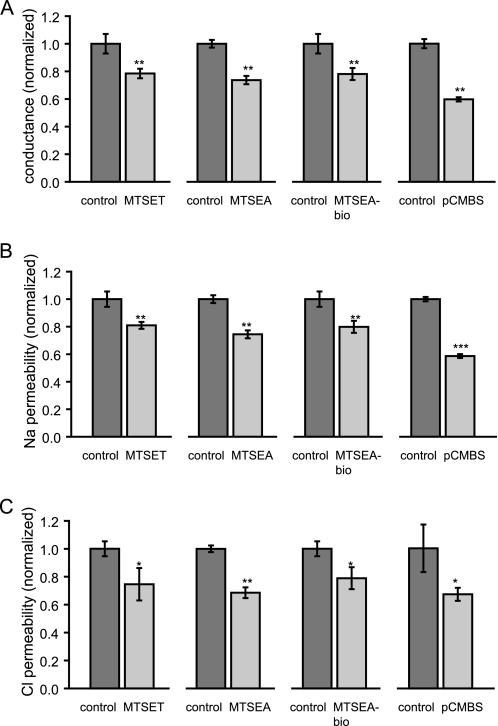
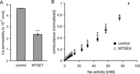
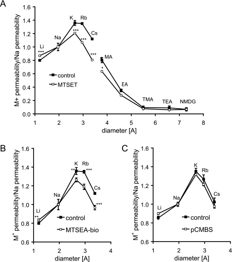
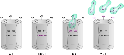
Claudins form size- and charge-selective pores in the tight junction that control the paracellular flux of inorganic ions and small molecules. However, the structural basis for ion selectivity of paracellular pores is poorly understood. Here we applied cysteine scanning to map the paracellular pathway of ion permeation across claudin-2-transfected Madin-Darby canine kidney type I cells. Four potential pore-lining amino acid residues in the first extracellular loop were mutated to cysteine and screened for their accessibility to thiol-reactive reagents. All mutants were functional except D65C, which formed dimers by intermolecular disulfide bonding, leading to a loss of charge and size selectivity. This suggests that claudin-2 pores are multimeric and that Asp(65) lies close to a protein-protein interface. Methanethiosulfonate reagents of different size and charge and the organic mercury derivate, p-(chloromercuri)benzenesulfonic acid, significantly decreased paracellular ion permeation across I66C-transfected cells by a mechanism that suggests steric blocking of the pore. The conductance of wild-type claudin-2 and the other cysteine mutants was only weakly affected. The rate of reaction with I66C decreased dramatically with increasing size of the reagent, suggesting that Ile(66) is buried deep within a narrow segment of the pore with its side group facing into the lumen. Furthermore, labeling with N-biotinoylaminoethyl methanethiosulfonate showed that I66C was weakly reactive, whereas Y35C was strongly reactive, suggesting that Tyr(35) is located at the protein surface outside of the pore.


Figures

FIGURE 1.
FIGURE 1.
Characterization of MDCK I TetOff cells, stably transfected with WT claudin-2 and the cysteine mutants Y35C, H57C, D65C, and I66C. The expression and distribution of claudin-2 was tested by double immunofluorescence staining with antibodies to claudin-2 and to the tight junction marker, ZO-1. Confocal en face images of induced cells (Dox) show that WT and mutant claudin-2 colocalize predominantly with ZO-1 to the tight junction but are also found in the cell bodies and at the apical membrane and, to a lesser extent, along the basolateral membrane (see also supplemental Fig. S1). Scale bar, 10 μm.
FIGURE 2.
FIGURE 2.
Western blot analysis of claudin-2 in cell lysates isolated from cell lines expressing WT claudin-2 and cysteine mutants. A, claudin-2 appears as a band at ∼22 kDa in induced cells (Dox), whereas expression is suppressed in the presence of doxycycline (Dox+). An additional band at between 40 and 45 kDa was observed with cells induced to express the D65C mutant of claudin-2. (The band at ∼35 kDa present in all lanes represents nonspecific antibody binding.) All samples were prepared with reducing sample buffer, containing 1% (v/v) 2-mercaptoethanol. B, testing the identity of the high molecular mass band at 40–45 kDa. Lysates of D65C and WT cells without pretreatment were prepared for SDS-PAGE using non-reducing sample buffer (lane 1) or reducing sample buffer (lane 2). Alternatively, intact cell monolayers were pretreated with 10 mm dithiothreitol (lane 3) or 110 mm Cu(II)-phenanthroline (lane 4) for 5 min to reduce and oxidize cysteines, respectively. The reagents were vigorously washed off prior to cell lysis, and samples were prepared with non-reducing buffer. The blot shows that preparation of the sample under reducing conditions or pretreatment of the cells with reducing reagent decreases the high molecular mass band.
FIGURE 3.
FIGURE 3.
Characterization of the electrophysiological properties of claudin-2 by Ussing chamber measurements of conductance and the transepithelial NaCl dilution potential. The figure shows representative data from two independent clones of MDCK I TetOff cells for each WT or cysteine mutant claudin-2. The results were corrected for the base-line conductance and permeability obtained with uninduced (Dox+) cells and represent the macroscopic conductance and permeability of claudin-2 pores. A, conductance of MDCK I TetOff cells induced to express WT and mutant claudin-2. The conductance of uninduced cells was typically <1 millisiemens (not shown). B, Na+ permeability (PNa). C, Cl permeability (PCl) of claudin-2 pores, calculated from the dilution potential. D, permeability ratio of Na+ to Cl (PNa/PCl). The diagram shows that D65C is much less cation-selective than WT claudin-2 and the other cysteine mutants. Data points represent means of 3–4 filters ± S.E. p values refer to statistically significant differences in comparison with WT claudin-2. *, p < 0.05; **, p < 0.01; ***, p < 0.001.
FIGURE 4.
FIGURE 4.
Ussing chamber measurements of bi-ionic diffusion potential to determine the permeabilities of WT and mutant claudin-2 for alkali metal ions (Eisenman sequence) and nitrogenous cations. A, the permeabilities of WT claudin-2, Y35C, H57C, and I66C to alkali metal ions are plotted as relative data compared with Na+ permeability. B, relative permeabilities of D65C and D65N to alkali metal ions in comparison with WT claudin-2, showing an increase in the relative permeabilities of D65C to larger cations. C, relative permeabilities of the nitrogenous cations, methylamine (MA), ethylamine (EA), tetramethylammonium (TMA), tetraethylammonium (TEA), and N-methyl-d-glucamine (NMDG). All were increased for D65C compared with WT claudin-2. The permeabilities of MA, EA, and TMA were fitted to a Renkin equation by non-linear regression to obtain the pore diameter, which was found to be as follow (±S.E.): 7.0 ± 0.8 Å (WT), 6.9 ± 0.1 Å (Y35C), 6.7 ± 0.1 Å (H57C), 10.2 ± 1.2 Å (D65C), 6.6 ± 0.3 Å (I66C). Data points represent means of four filters ± S.E. *, p < 0.05; **, p < 0.01; ***, p < 0.001. p values refer to statistically significant differences in comparison with WT claudin-2.
FIGURE 5.
FIGURE 5.
Time-dependent measurements of conductance of WT and mutant claudin-2 and effects of thiol-reactive reagents. MTS and pCMBS were added to both sides of the cell monolayers mounted in Ussing chambers. The graphs show representative data of induced and uninduced cells incubated with MTSET (1 mm) (A), MTSEA (2.5 mm) (B), MTSES (5 mm, only WT and I66C shown) (C), and pCMBS (0.5 mm, only WT and I66C shown) (D). In all measurements, the effects on uninduced cells were very small.
FIGURE 6.
FIGURE 6.
A, quantification of SCAM conductance measurements. The diagram shows the relative changes in conductance of WT and mutant claudin-2 induced by different MTS reagents. The data are means of three filters ± S.E. of representative experiments, measured 3 min after the addition of MTS. B, testing the reaction of MTSES and MTSACE with I66C. Cells expressing I66C were preincubated with either MTSES or MTSACE for 5 min prior to the addition of MTSEA. Control cells were not preincubated. Preincubation with either MTSES or MTSACE largely prevented the inhibition of conductance by MTSEA. C, Western blot of claudin-2 to test the reaction of endogenous and engineered cysteines with MTSEA-biotin. Intact cell layers were incubated with MTSEA-biotin, and cells were homogenized with radioimmune precipitation buffer. Samples were taken from the whole cell lysate to test overall claudin-2 expression (bottom). Biotin-labeled proteins were affinity-purified with streptavidin-coated agarose beads, and biotinylated claudin-2 was detected using a claudin-2-specific antibody (top).
FIGURE 7.
FIGURE 7.
Kinetics of the inhibition of conductance of I66C by different MTS reagents. Cationic MTS reagents of increasing size, MTSEA, MTSET, and MTSPTrEA, and the uncharged reagent, MTSEA-biotin, were used. A, raw data showing change in normalized conductance with time after adding the MTS reagent. B, relationship between the molecular mass of the reagent and the second order rate constant, k.
FIGURE 8.
FIGURE 8.
Ussing chamber experiments with I66C to determine the effect of thiol-reactive reagents of different size and charge, MTSEA, MTSET, and MTSEA-biotin pCMBS, on conductance (A), Na+ permeability (PNa) (B), and Cl permeability (PCl) (C) of claudin-2 pores. The MTS- and pCMBS-induced decreases in conductance correlate with similar changes in PNa and PCl. Data points represent means of four filters ± S.E. The data are corrected for measurements with uninduced cells. *, p < 0.05; **, p < 0.01; ***, p < 0.001. p values refer to statistically significant differences compared with control cells.
FIGURE 9.
FIGURE 9.
Testing electrostatic effects created by positively charged MTS after reaction with I66C. A, measurements of Ca2+ permeability (PCa) of control and MTSET-treated cells, grown on Transwell filters, by tracer flux experiments (n = 4). B, to unmask surface charge effects, the dependence of conductance on ionic strength was determined in Ussing chambers. The NaCl activity of the medium of control and MTSEA-treated cells was gradually increased from 0 to 90 mm, replacing with mannitol as needed to maintain osmolality (n = 3). ***, p < 0.001, compared with control.
FIGURE 10.
FIGURE 10.
Bi-ionic diffusion potential measurements to determine the effect of different MTS reagents and pCMBS on the relative cation permeability properties of I66C. The data were plotted as relative data compared with PNa. A, effect of MTSET on the relative permeabilities of alkali metal cations (Eisenman sequence) and nitrogenous cations of different diameter. Shown are the effects of MTSEA-biotin (B) and pCMBS (C) on the Eisenman sequence of I66C. Data points represent means of four filters ± S.E. *, p < 0.05; **, p < 0.01; ***, p < 0.001. p values refer to statistically significant differences compared with untreated control cells.
FIGURE 11.
FIGURE 11.
Model of claudin-2 showing the putative location of the mutated residues in the first extracellular domain. The pore is hypothesized to be a homomultimer (residues from 2–3 subunits are shown). Wild-type claudin-2 (WT) is depicted in the left panel, and the consequences of cysteine mutagenesis are shown in the other panels. D65 is located in the narrowest part of the pore facing the lumen and close to an intersubunit interface, so that the D65C mutation leads to dimerization by disulfide bonding. Ile66 is also within the pore facing the lumen, but residues from neighboring subunits are further apart, so that disulfide bonding in I66C is precluded. Thiol-reactive MTS reagents (green) enter the pore to react with I66C, partially blocking the pore to ion permeation. Only a single MTS molecule is accommodated in each pore. Tyr35 is outside the pore facing extracellularly. Thus, reaction of MTS reagents with Y35C does not block the pore. Furthermore, because there is no steric restriction, every Y35C residue can react with an MTS molecule, so that multiple MTS molecules are associated with each pore multimer.

References

    1. Furuse M., Fujita K., Hiiragi T., Fujimoto K., Tsukita S. (1998) J. Cell Biol. 141, 1539–1550 - PMC - PubMed
    1. Tsukita S., Furuse M. (2000) J. Cell Biol. 149, 13–16 - PMC - PubMed
    1. Van Itallie C. M., Anderson J. M. (2006) Annu. Rev. Physiol. 68, 403–429 - PubMed
    1. Van Itallie C. M., Holmes J., Bridges A., Gookin J. L., Coccaro M. R., Proctor W., Colegio O. R., Anderson J. M. (2008) J. Cell Sci. 121, 298–305 - PubMed
    1. Yu A. S., Cheng M. H., Angelow S., Günzel D., Kanzawa S. A., Schneeberger E. E., Fromm M., Coalson R. D. (2009) J. Gen. Physiol. 133, 111–127 - PMC - PubMed

Publication types

LinkOut - more resources