. 2009 Sep;119(9):2601-12.
doi: 10.1172/JCI38323. Epub 2009 Aug 17.

Aldosterone mediates activation of the thiazide-sensitive Na-Cl cotransporter through an SGK1 and WNK4 signaling pathway

Affiliations

Aldosterone mediates activation of the thiazide-sensitive Na-Cl cotransporter through an SGK1 and WNK4 signaling pathway

David J Rozansky et al. J Clin Invest. 2009 Sep.

Abstract

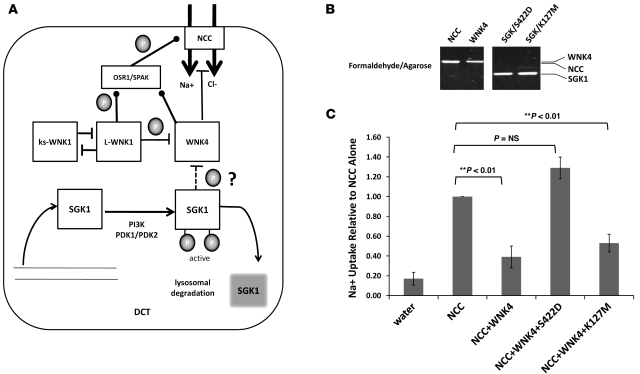
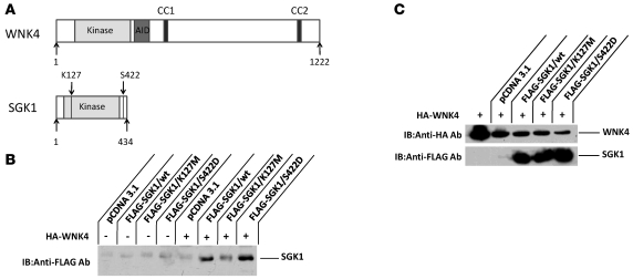
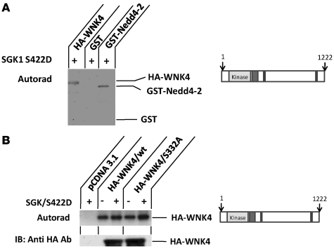
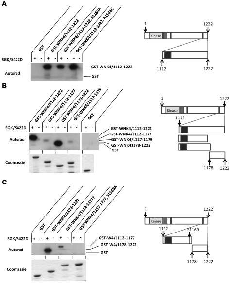
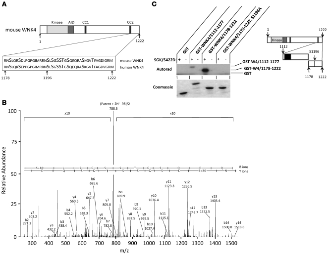
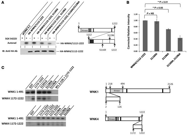
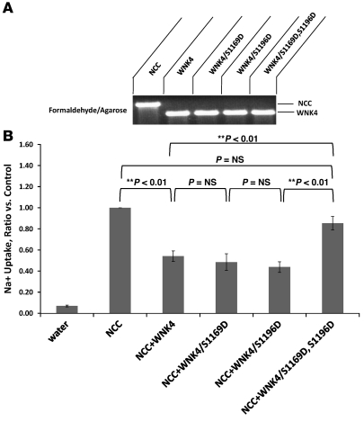
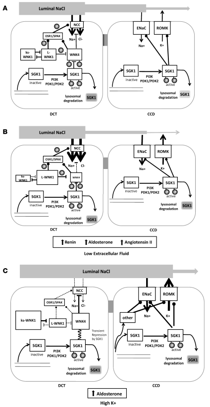
Aldosterone regulates volume homeostasis and blood pressure by enhancing sodium reabsorption in the kidney's distal nephron (DN). On the apical surface of these renal epithelia, aldosterone increases expression and activity of the thiazide-sensitive Na-Cl cotransporter (NCC) and the epithelial sodium channel (ENaC). While the cellular mechanisms by which aldosterone regulates ENaC have been well characterized, the molecular mechanisms that link aldosterone to NCC-mediated Na+/Cl- reabsorption remain elusive. The serine/threonine kinase with-no-lysine 4 (WNK4) has previously been shown to reduce cell surface expression of NCC. Here we measured sodium uptake in a Xenopus oocyte expression system and found that serum and glucocorticoid-induced kinase 1 (SGK1), an aldosterone-responsive gene expressed in the DN, attenuated the inhibitory effect of WNK4 on NCC activity. In addition, we showed--both in vitro and in a human kidney cell line--that SGK1 bound and phosphorylated WNK4. We found one serine located within an established SGK1 consensus target sequence, and the other within a motif that was, to our knowledge, previously uncharacterized. Mutation of these target serines to aspartate, in order to mimic phosphorylation, attenuated the effect of WNK4 on NCC activity in the Xenopus oocyte system. These data thus delineate what we believe to be a novel mechanism for aldosterone activation of NCC through SGK1 signaling of WNK4 kinase.


Figures

Figure 1
Figure 1. The active form of SGK1 reverses WNK4 inhibition of NCC-mediated Na+ uptake in Xenopus oocytes.
(A) Schematic diagram of the DCT, outlining the hypothesis of where SGK1 acts to influence WNK4 inhibition of Na+/Cl cotransport (shown by thick arrows). The question mark and dashed line indicate the area of interest for this study. Arrows denote SGK1 movement among inactive, active, and degraded forms. Stimulatory and inhibitory effects are indicated by filled circles and blunt-headed arrows, respectively. Phosphorylation steps are denoted by “P.” (B) Representative sample of formaldehyde/agarose gel stained with ethidium bromide showing equivalent amounts of SGK/S422D and SGK/K127M cRNA and no obvious degradation prior to injection into oocytes. Lanes were run on the same gel, which was split to maintain the sample order in C. (C) Relative to NCC alone, WNK4 reduced NCC-mediated Na+ flux by 60%. The addition of constitutively active SGK1/S422D reversed that effect, whereas addition of kinase-dead SGK1/K127M continued to reduce Na+ flux. n = 3 for each condition (± SEM). Significance (by ANOVA) is indicated.
Figure 2
Figure 2. SGK1 associates with WNK4.
(A) Schematic of WNK4, showing kinase domain, autoinhibitory domain (AID), and coiled-coil domains (CC), and of SGK1, showing kinase domain and K127M (kinase-dead) and S422D (constitutively active) mutation locations. (B) HEK293 cells were transfected with FLAG-tagged SGK1 isoforms or vector pCDNA3.1 in the presence or absence of full-length HA-tagged WNK4. After lysis and isolation with anti-HA sepharose beads, coimmunoprecipitation between SGK1 and WNK4 was tested by immunoblot using anti-FLAG antibody. The blot revealed the presence of SGK1 protein bound to WNK4. WNK4 associated with SGK1 with greater signal for wild-type and S422D isoforms than for the K127M isoform. The background band evident in all lanes is the IgG light chain, which migrated on the gel at a slightly larger molecular weight than SGK1. (C) Immunoblots from transfected cell lysates only, confirming the presence of WNK4 and SGK1 for interactions tested in B. Blots in B and C are representative samples from 2 experiments.
Figure 3
Figure 3. SGK1 kinase activity on full-length WNK4.
(A) SGK1 kinase assay testing HA-tagged WNK4, GST-Nedd4-2, and GST alone as substrates. WNK4 and GST-Nedd4-2, but not GST alone, were phosphorylated. (B) SGK1 kinase assay of HA-tagged WNK4 and autophosphorylation-deficient WNK4 (S332A). A stronger signal was noted in the presence of SGK1, but its absence did not eliminate the signal. The immunoblot below confirmed equivalent amounts of the 2 WNK4 isoforms used in the SGK1 kinase assay above. Blots are representative samples from 2 experiments.
Figure 4
Figure 4. SGK1 phosphorylates more than 1 serine/threonine site of the C-terminal region of WNK4.
GST-WNK4 fusion proteins from 1112 to 1222 were isolated from bacteria and subjected to an SGK1 kinase assay. Shown with each panel is a schematic of the WNK4 region of interest and the approximate size of the GST-fusion protein. (A) SGK1 kinase assay of 0.8 μg GST-WNK/1112–1222 and isoforms conforming to mutants associated with FHHt (R1164C) and putative SGK1 phosphorylation target (S1169A). Autoradiographic signal was equivalent for wild-type and R1164C, with a slight reduction in signal for S1169A. (B) C-terminal GST-WNK4 deletion constructs showed much stronger signal for 1178–1222 compared with 1112–1177 or 1127–1179. Below, Coomassie Blue staining of protein preparations confirmed that equivalent amounts of the 5 substrates were used in the study above. Data are from 2 gels run simultaneously. (C) S1169 was confirmed as the only target of SGK1 phosphorylation in WNK4 fragment 1112–1177. Shown below is Coomassie Blue staining of 4 protein preparations. Blots in AC are representative samples from 3 experiments.
Figure 5
Figure 5. Within the 45–amino acid C-terminal NCC negative regulatory region of WNK4/1178–1222, S1196 acts as a single target of SGK1 phosphorylation.
(A) Amino acid sequence of this WNK4 region contains 10 serines and threonines. (B) MS/MS spectrum of peptide 246–260 from a tryptic digest of GST-WNK4/1178–1222, localizing phosphorylation to residue S248 in the construct (corresponding to residue S1196 of mouse WNK4) after incubation with SGK1 kinase. An uninterrupted y and b ion series was observed in the fragment ion spectra, as well as a characteristic major doubly charged neutral loss ion at m/z 788.5 resulting from the loss of phosphate. Because of the intensity of this neutral loss ion, the intensities of the other areas of the spectrum increased 10-fold. The inset sequence of the peptide is aligned with the corresponding fragment ions of the y and b ion series. (C) In vitro SGK1 kinase assay confirmed that S1196 was the only SGK1 phosphorylation site in the region 1178–1222 from bacterially derived fusion fragments. The single mutation of WNK4/1178–1222 S1196A was not phosphorylated by SGK1, whereas wild-type and fragment 1112–1177 yielded robust and weak signals, respectively. Shown below is Coomassie Blue staining of the 4 protein preparations used for the SGK kinase assay. n = 3. The schematic diagram denotes the C-terminal WNK4 region and the approximate size of the GST-fusion proteins.
Figure 6
Figure 6. SGK1 and WNK1 phosphorylate S1196 of the C-terminal fragment of WNK4 kinase.
(A) HA-tagged WNK4/1112–1222 was mutated at S1169 and/or S1196 and isolated by immunoprecipitation from HEK293 cells. In vitro SGK1 kinase assay of these fragment isoforms showed that wild-type and S1169A had higher levels of autoradiographic signal compared with S1196A or the double mutant S1169A/S1196A. Immunoblot shown below demonstrates comparable protein levels of the 4 fragments used above. n = 3. (B) Average ratios of autoradiographic densitometric signal to immunoblot signal for each fragment, which revealed additive properties with reduction in signal as index serines were removed. While S1169A trended lower, significance in signal reduction was noted for S1196A and the double mutant. Significance (by ANOVA) is shown. n = 3 for each condition (± SEM). (C) Schematic diagram shows the constructs used in a WNK1 kinase assay for both WNK1 and WNK4. The top blot shows WNK1 kinase assays of GST-WNK1 fusion enzymes in the presence of substrates: GST alone, or GST-WNK4 C-terminal fragment 1172–1222 and specific serine-to-alanine mutations. The first row demonstrated that GST-WNK1/1–491 had autophosphorylation properties, whereas GST-WNK1/2–126 did not. The second row indicated that C-terminal serine-to-alanine mutations of WNK4 reduced the phosphorylation signal for WNK4/S1196 exclusively. In the bottom blot, the first row replicates indicated equal autophosphorylation of WNK1. In the second row, the WNK4/S1196A substrates had a weaker signal than that of their wild-type counterparts.
Figure 7
Figure 7. The double aspartate WNK4 mutant S1169D,S1196D fails to inhibit NCC activity in Xenopus oocytes.
(A) Formaldehyde/agarose gel stained with ethidium bromide showed equivalent amounts of cRNA and no obvious degradation for each WNK4 construct prior to oocyte injections. (B) Relative to NCC alone, WNK4 reduced NCC-mediated Na+ flux by 50%. Single aspartate mutations at either S1169 or S1196 did not significantly impair the ability of WNK4 to block NCC activity. However, WNK4 containing both serine-to-aspartate mutations was unable to reduce Na+ uptake. n = 3 for each condition (± SEM). Significance (by ANOVA) is shown.
Figure 8
Figure 8. WNK4, WNK1, SGK1, and aldosterone paradox hypothesis.
WNK4 kinase inhibits NCC activity. Multiple secondary messengers modulate WNK4 action. L-WNK1 inhibits WNK4, but is suppressed by ks-WNK1. L-WNK1 also phosphorylates OSR1/SPAK, thereby activating NCC. Symbols are as in Figure 1; secondary messenger proteins and ion transporter sizes vary relative to their proposed activity. (A) Our results suggest that SGK1, induced transcriptionally by aldosterone and activated by PI3K, negatively regulates WNK4’s action on NCC by phosphorylating 2 C-terminal serines of WNK4. SGK1 also positively effects ENaC and ROMK activity in the cortical collecting duct (CCD) through regulatory pathways that include SGK1. (B) Schematic proposal of renal response to hypovolemia, through hyperreninemia and/or stimulation of angiotensin II and aldosterone. Here SGK1 and WNK1 target similar C-terminal WNK4 serines for phosphorylation, relieving the WNK4 inhibitory effect on NCC, increasing NCC-mediated Na+/Cl cotransport (shown by arrow thickness) while diminishing the regulatory value of SGK1 and other mechanisms in the CCD that lead to K+ secretion. (C) Schematic proposal of renal response to aldosterone stimulation by hyperkalemia. In the DCT there is upregulation of ks-WNK1 activity, which — by squelching L-WNK1 — leads to heightened WNK4 activity, low SPAK/OSR1 activity, and reduced NCC-mediated NaCl transport. We surmise that SGK1 has a transient effect on WNK4 with downstream ENaC and ROMK activation by SGK1 and other aldosterone-sensitive mechanisms, restoring potassium balance without affecting extracellular volume.

References

    1. Reilly R.F., Ellison D.H. Mammalian distal tubule: physiology, pathophysiology, and molecular anatomy. Physiol. Rev. 2000;80:277–313. - PubMed
    1. Pearce D., Kleyman T.R. Salt, sodium channels, and SGK1. J. Clin. Invest. 2007;117:592–595. doi: 10.1172/JCI31538. - DOI - PMC - PubMed
    1. Verrey F., Loffing J., Zecevic M., Heitzmann D., Staub O. SGK1: aldosterone-induced relay of Na+ transport regulation in distal kidney nephron cells. Cell. Physiol. Biochem. 2003;13:21–28. doi: 10.1159/000070246. - DOI - PubMed
    1. Lang F., et al. (Patho)physiological significance of the serum- and glucocorticoid-inducible kinase isoforms. Physiol. Rev. 2006;86:1151–1178. doi: 10.1152/physrev.00050.2005. - DOI - PubMed
    1. von Wowern F., et al. Genetic variance of SGK-1 is associated with blood pressure, blood pressure change over time and strength of the insulin-diastolic blood pressure relationship. Kidney Int. 2005;68:2164–2172. doi: 10.1111/j.1523-1755.2005.00672.x. - DOI - PubMed

Publication types

MeSH terms