. 2009 Sep;119(9):2725-36.
doi: 10.1172/JCI39542. Epub 2009 Aug 17.

Binding of pro-prion to filamin A disrupts cytoskeleton and correlates with poor prognosis in pancreatic cancer

Affiliations

Binding of pro-prion to filamin A disrupts cytoskeleton and correlates with poor prognosis in pancreatic cancer

Chaoyang Li et al. J Clin Invest. 2009 Sep.

Abstract

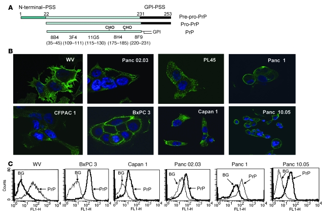
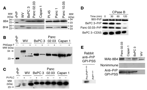
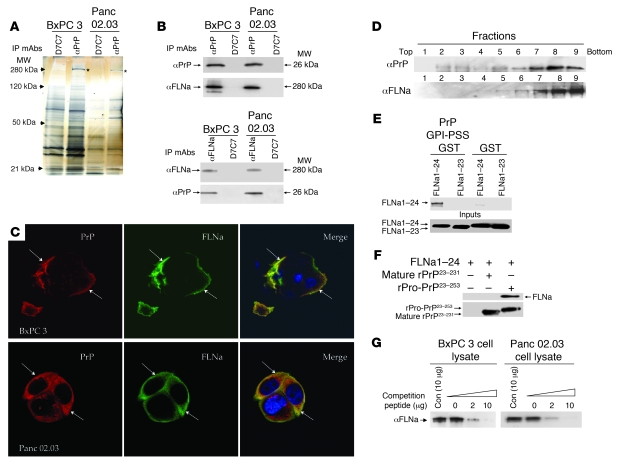
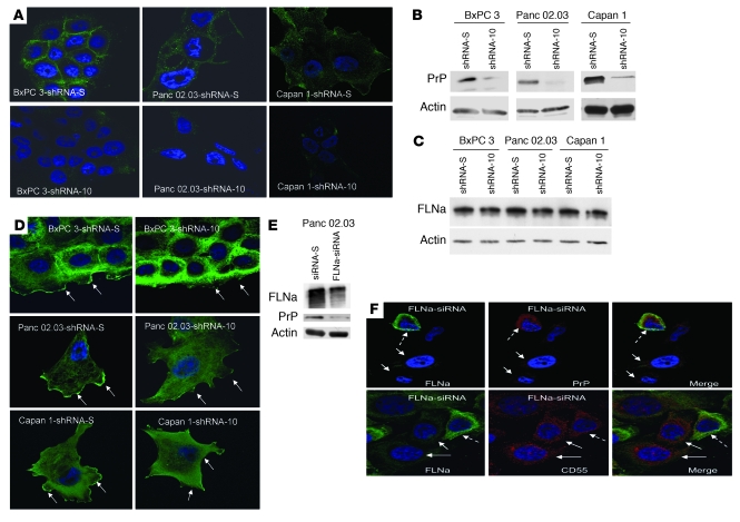
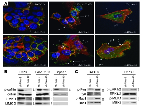
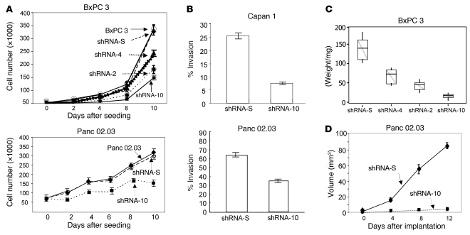
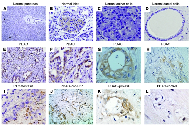
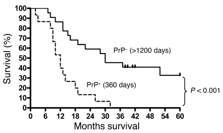
The cellular prion protein (PrP) is a highly conserved, widely expressed, glycosylphosphatidylinositol-anchored (GPI-anchored) cell surface glycoprotein. Since its discovery, most studies on PrP have focused on its role in neurodegenerative prion diseases, whereas its function outside the nervous system remains unclear. Here, we report that human pancreatic ductal adenocarcinoma (PDAC) cell lines expressed PrP. However, the PrP was neither glycosylated nor GPI-anchored, existing as pro-PrP and retaining its GPI anchor peptide signal sequence (GPI-PSS). We also showed that the PrP GPI-PSS has a filamin A-binding (FLNa-binding) motif and interacted with FLNa, an actin-associated protein that integrates cell mechanics and signaling. Binding of pro-PrP to FLNa disrupted cytoskeletal organization. Inhibition of PrP expression by shRNA in the PDAC cell lines altered the cytoskeleton and expression of multiple signaling proteins; it also reduced cellular proliferation and invasiveness in vitro as well as tumor growth in vivo. A subgroup of human patients with pancreatic cancer was found to have tumors that expressed pro-PrP. Most importantly, PrP expression in tumors correlated with a marked decrease in patient survival. We propose that binding of pro-PrP to FLNa perturbs FLNa function, thus contributing to the aggressiveness of PDAC. Prevention of this interaction could provide an attractive target for therapeutic intervention in human PDAC.


Figures

Figure 1
Figure 1. Expression of PrP in the PDAC cell lines exists as pro-PrP.
(A) A diagram of the processing of GPI-anchored PrP and the epitopes of the mAbs (CHO, N-linked glycans). (B) Confocal microscopic images show that WV cells express PrP on the cell surface. All 7 PDAC cell lines express varying levels of PrP on the cell surface as well as in the cytoplasm. Original magnification, ×1,000. (C) Histograms show the presence of PrP on the cell surface of live PDAC cell lines. BG, background, cells stained with control irrelevant mAb D7C7.
Figure 2
Figure 2. PrP in the PDAC cell lines exists as pro-PrP.
(A) Immunoblots show PrP from WV cells has a MW of 34 kDa, while PrP from the PDAC cell lines has a MW of 26 kDa. A recombinant PrP (rPrP) produced in E. coli is included as a control and MW marker. (B) Immunoblots show treatment of PrP from WV cells with endoglycosidase-F (PNGase F) reduces its MW from 34 kDa to 25.5 kDa. But identical treatment does not change the mobility of PrP from the PDAC cell lines. Deglycosylated PrP from WV cells migrated slightly faster than PrP from the PDAC cell lines (dashed arrows). (C) Immunoblots show PrP from WV cells is sensitive to PI-PLC, as shown by the appearance of a smaller PrP species (bottom arrow) in addition to the PNGase F–treated species (top arrow), but PrP from the PDAC cell lines is resistant to PI-PLC. (D) Immunoblots show that while PrP from the 2 PDAC cell lines is sensitive to CPase B, PrP from WV cells is resistant. CD55 from BxPC 3 cells is also resistant to CPase B. (E) Immunoblots show a rabbit antiserum specific for the PrP GPI-PSS reacts with recombinant pro-PrP (rPro-PrP23–253) but not with recombinant mature PrP (rPrP23–231). The anti–GPI-PSS antiserum also reacts with pro-PrP from the PDAC cell lines but does not react with the PrP from WV cells.
Figure 3
Figure 3. FLNa binds to the GPI-PSS of pro-PrP.
(A) A silver-stained gel shows a band with MW of 280 kDa (*) is coimmunoprecipitated with anti-PrP mAb 8B4 but not with control mAb D7C7. (B) Immunoblots show the copurification of FLNa with PrP and vice versa. (C) Confocal microscopic images show colocalization of FLNa (green) and PrP (red) in PDAC cell lines. Arrows show area of colocalization. Original magnification, ×1,000. (D) Immunoblots show PrP and FLNa are present in similar fractions after centrifugation in sucrose gradient. (E) An in vitro pull-down experiment shows much stronger binding of full-length FLNa1–24 to a PrP GPI-PSS GST fusion protein than to a GST protein without the GPI-PSS. A FLNa1–23 monomer did not bind PrP GPI-PSS GST fusion protein. Immune complexes were pulled down with GST binding beads and immunoblotted with an anti-FLAG mAb to detect FLNa. (F) Immunoblots show binding of FLNa1–24 to recombinant pro-PrP23–253 but not mature recombinant PrP23–231. Anti-PrP mAb 8H4 was used to pull down the immune complexes. The immunoblot was done either with an anti-Flag mAb or anti-PrP mAb 8H4. (G) Immunoblots show competition of binding of FLNa to pro-PrP by a PrP-GPI-PSS synthetic peptide. Copurification of PrP and FLNa in the PDAC cell lysates was carried out in the presence of different concentrations of either a synthetic peptide corresponding to the GPI-PSS or a control synthetic peptide. Anti-PrP mAb 8B4 coimmunoprecipitated proteins were then immunoblotted with an anti-FLNa mAb. Con, control peptide.
Figure 4
Figure 4. Downregulation of PrP or FLNa expression in the PDAC cell lines.
(A) Immunofluorescence staining and confocal microscopic images show the PDAC cell lines with shRNA-10 have reduced levels of PrP. Original magnification, ×1,000. (B) Immunoblots show the PrP-downregulated shRNA-10 cells have reduced levels of PrP. (C) Immunoblots show the level of FLNa does not change in PrP-downregulated cells. (D) Immunofluorescence staining and confocal microscopic images show that knocking down PrP alters the spatial distribution of FLNa. Arrows show staining of membrane ruffles and leading edges. Original magnification, ×1,000. (E) Immunoblots show that when expression of FLNa is inhibited, the expression of PrP is also reduced in Panc 02.03 cells. (F) Immunofluorescence staining and confocal microscopic images show the expression of FLNa modulates PrP but not CD55 expression. In the top left panel, the dashed arrow identifies a cell with FLNa (green); solid arrows identify 2 cells lacking FLNa. In the top center panel, 2 solid arrows identify 2 cells lacking PrP; the dashed arrow identifies 1 cell with PrP (red). The top right panel is the merge of the left and center panels; 2 arrows identify 2 cells lacking both PrP and FLNa, and a dashed arrow identifies 1 cell with both PrP and FLNa. In the bottom panels, 2 FLNa-negative cells (left panel, solid arrows) still express high levels of CD55 (red; center panel, solid arrows), although some cells have both FLNa and CD55 stain (left and center panels, dashed arrows). Original magnification, ×1,000.
Figure 5
Figure 5. Binding of pro-PrP to FLNa alters actin organ­ization and signaling events.
(A) Immuno­fluorescence staining and confocal microscopic images show that knocking down PrP modifies the spatial distribution of actin filaments (red) and p-Tyr (green) in 3 PDAC cell lines. (a, actin, shown with dashed arrows; p-T, p-Tyr, shown with solid arrows). Original magnification, ×1,000. (B) Immunoblots of PrP-downregulated BxPC 3 and Panc 02.03 cells show that the levels of p-cofilin, LIMK1, and LIMK2 are markedly reduced (open arrows, the size of the arrow indicates the degree of change) compared with control cells. P-cofilin is also reduced in PrP-downregulated Capan 1 cells. (C) Immunoblots show upregulation of p-Fyn, p-Rac1, p-ERK1/2, and p-MEK1 (open arrows) in PrP-downregulated BxPC 3 cells.
Figure 6
Figure 6. Downregulation of PrP influences the in vitro and in vivo behavior of the PDAC cell lines.
(A) Proliferation of PrP-downregulated cells is reduced compared with control cells with scrambled shRNA-S or cells without any shRNA. Parental BxPC 3 cells (open circles); BxPC 3–shRNA-S cells (filled diamonds); BxPC 3–shRNA-4 cells (filled triangles); BxPC 3–shRNA-2 cells (filled circles); BxPC 3–shRNA-10 cells (filled squares); parental Panc 02.03 cells (open circles); Panc 02.03–shRNA-S cells (filled diamonds); Panc 02.03–shRNA-10 cells (filled squares). The results presented are the mean of triplicate wells ± SD. (B) In vitro invasiveness of PrP-downregulated Capan 1–shRNA-10 cells and Panc 02.03–shRNA-10 cells in Matrigel is reduced. The results presented are the mean of triplicate wells ± SD. (C) In vivo growth of PrP-downregulated BxPC 3 cells in nude mice depends on the levels of PrP expression (n = 10/group, composite of 2 experiments; n = 5/experiment). The upper whisker extends to the highest value within the upper limit. The lower whisker extends to the lowest value within the lower limit. The top of the box is the third quartile. The bottom of the box is the first quartile. The median of the data is also indicated by the horizontal line. (D) The growth of PrP-downregulated Panc 02.03–shRNA-10 cells in nude mice is inhibited. Panc 02.03–shRNA-S cells (filled diamonds); Panc 02.03–shRNA-10 cells (filled squares). The results presented are the mean of 10 mice/group ± SD (composite of 2 experiments, n = 5/experiment).
Figure 7
Figure 7. PrP is present in PDAC lesions but not in normal ductal cells.
Immunohistochemical staining shows that in normal pancreas (AD) only islet cells express PrP. (A) Two arrows identify 2 islets (original magnification, ×100). (B) A PrP-positive islet (original magnification, ×400). (C) Neither acinar cells, an arrow shows a centroacinar cell (original magnification, ×400), nor (D) ductal cells (original magnification, ×400) express PrP. (EH) In PDAC, malignant ductal cells express PrP (original magnification, ×200 [E]; ×400 [H]). F and G are from 2 additional PDAC patients (original magnification, ×400). (G) The dashed arrow shows immunoreactivity on the cell surface. (I) PDAC lymph node metastases express PrP (original magnification, ×400). (J) PrP in PDAC reacted with the anti–PrP-GPI-PSS antibody; 3 arrows identify tumors (original magnification, ×200). (K) Dashed arrows in K indicate PDAC cell surface immunoreactivity (original magnification, ×400). (L) The control antiserum only has background immunoreactivity (original magnification, ×400).
Figure 8
Figure 8. Expression of PrP is associated with poorer prognosis.
The 37 patients had surgery done from 2001 to 2003. Patients (n = 16) whose tumors expressed PrP (PrP+) had a median survival time of 360 days. On the other hand, of the 21 patients whose tumors lacked PrP (PrP), 6 of these patients are still alive as of October of 2008. Four of these patients have already passed 5 years after surgery; 2 others will have passed 5 years in late November of 2008 (2 of the spikes). The other 2 spikes indicate a patient who died 41 months after surgery and a patient who died 52 months after surgery, respectively. This cohort of patients has mean survival time of more than 1,200 days (P < 0.001).

Similar articles

Cited by

References

    1. Prusiner S.B. Prions. Proc. Natl. Acad. Sci. U. S. A. 1998;95:13363–13383. doi: 10.1073/pnas.95.23.13363. - DOI - PMC - PubMed
    1. Brockes J.P. Topics in prion cell biology. Curr. Opin. Neurobiol. 1999;9:571–577. doi: 10.1016/S0959-4388(99)00016-1. - DOI - PubMed
    1. Hegde R.S., Rane N.S. Prion protein trafficking and the development of neurodegeneration. Trends Neurosci. . 2003;26:337–339. doi: 10.1016/S0166-2236(03)00143-7. - DOI - PubMed
    1. Mouillet-Richard S., et al. Signal transduction through prion protein. Science. 2000;289:1925–1928. doi: 10.1126/science.289.5486.1925. - DOI - PubMed
    1. Taylor D.R., Hooper N.M. The prion protein and lipid rafts. Mol. Membr. Biol. 2006;23:89–99. doi: 10.1080/09687860500449994. - DOI - PubMed

Publication types

MeSH terms