. 2009 Jul-Aug;3(4):233-8.
doi: 10.4161/chan.3.4.9381. Epub 2009 Jul 24.

Synaptic NMDAR activity suppresses FOXO1 expression via a cis-acting FOXO binding site: FOXO1 is a FOXO target gene

Affiliations

Synaptic NMDAR activity suppresses FOXO1 expression via a cis-acting FOXO binding site: FOXO1 is a FOXO target gene

Bashayer Al-Mubarak et al. Channels (Austin). 2009 Jul-Aug.

Abstract

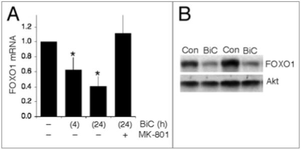
Activation of gene expression by FOXO transcription factors can promote neuronal death in response to loss of trophic support, or oxidative stress. The predominant neuronal FOXOs, FOXO1 and FOXO3, promote the expression of pro-death genes, such as Fas Ligand, Bim and Txnip. Neuroprotective signals initiated by neurotrophins, growth factors or synaptic activity trigger the nuclear export of FOXOs via activation of the PI3K-Akt pathway. One key aspect of FOXO regulation is that once PI3K-Akt activity has returned to baseline, FOXOs return to the nucleus to resume the activation of their target genes. Thus, the FOXO-inhibiting capacity of the PI3K-Akt pathway is thought to be short-lived. However, we show here that synaptic NMDA receptor activity not only triggers FOXO export, but also suppresses the expression of FOXO1. Blockade of PI3K activity prevents both FOXO nuclear export and suppression of FOXO1 expression, raising the possibility that FOXO1 is itself a FOXO target gene. We found that FOXO3, and to a lesser extent FOXO1 transactivates the FOXO1 promoter via a consensus FOXO binding site (GTA AAC AA), and also an upstream sequence resembling a classical FOXO-binding insulin response sequence (CAA AAC AA). Activity-dependent suppression of the FOXO1 promoter is mediated through the proximal GTAAACAA sequence. Similar suppression via this site is observed by activating neuronal IGF-1 receptors by exogenous insulin. Thus, through a feed-forward inhibition mechanism, synaptic activity triggers FOXO export resulting in suppression of FOXO1 expression. These results suggest that FOXO-inactivating signals are likely to result in longer-term inhibition of FOXO target gene expression than previously thought.


Figures

Figure 1
Figure 1
Synaptic nMdA receptor activity suppresses expression of FOXO1. (A) Qrt-PCr analysis of FOXO1 mrnA levels in neurons placed in trophically deprived medium and stimulated as indicated. Levels of FOXO1 mrnA are normalized to those of GAPdH. *p < 0.05 (AnOVA followed by Fisher’s LSd test here and throughout), n = 5. (B) Example western blot demonstrating that activity-dependent suppression of FOXO1 mrnA levels results in lower protein expression. Two sets of protein lysates are shown, taken from independent cultures, to illustrate the effect of BiC/4-AP stimulation on FOXO1 protein levels after 24 h. For comparison, Akt levels are shown to be similar.
Figure 2
Figure 2
Activity-induced FOXO export and suppression of FOXO1 expression is Pi3K-dependent. (A) Schematic illustration of the position and conservation of the putative FOXO-response elements within the FOXO1 promoter. Nucleotide positions refer to the rat gene. (B) Neuronal activity promotes the nuclear export of FOXO3 via Pi3K. Neurons were transfected with an expression vector encoding myc-tagged FOXO3, plus a peGFP transfection marker. The neurons were then stimulated with BiC/4-AP as indicated for 1 h in the presence or absence of the Pi3K inhibitor LY294002 (50 μM). The cells were then fixed and processed for immunofluorescence with an anti-myc antibody as described previously. Representative pictures are shown from the experiments performed. White arrows indicate transfected cells. (C) Qrt-PCr analysis of FOXO1 mrnA levels in neurons placed in trophically deprived medium and stimulated as indicated in the presence or absence of the Pi3K inhibitor LY294002 (50 μM). *p < 0.05 (n = 3).
Figure 3
Figure 3
FOXO consensus sites mediate activity-dependent suppression and FOXO-mediated transactivation of the FOXO1 promoter. (A) Neurons were transfected with either FOXO1-Luc or FOXO1(mut1)-Luc, plus a pTK-renilla normalization vector. Neurons were stimulated where indicated with BiC/4-AP or placed into trophic medium containing insulin, for 24 h, after which reporter activity levels were measured (normalized to renilla levels). *p < 0.05 (n = 4). (B) Neurons were transfected with either FOXO1-Luc or FOXO1(mut1)-Luc, plus a pTK-renilla normalization vector. In addition, they were cotransfected with a vector encoding FOXO1, FOXO3, or beta-globin (control). Reporter activity levels were measured (normalized to renilla levels) at 48 h post-transfection. *p < 0.05 assessment of the effect of FOXO expression relative to appropriate control, #p < 0.05 assessment of the effect of the mutation on basal, and FOXO-induced activity of the FOXO1 promoter (n = 7). (C) Neurons were transfected with either FOXO1-Luc or mutated variants, plus a pTK-renilla normalization vector. In addition, they were cotransfected with a vector encoding either FOXO3, or beta-globin (control). Reporter activity levels were measured (normalized to renilla levels) at 48 h post-transfection. *p < 0.05 (n = 5).

References

    1. Mariani E, Polidori MC, Cherubini A, Mecocci P. Oxidative stress in brain aging, neurodegenerative and vascular diseases: an overview. J Chromatogr B Analyt Technol Biomed Life Sci. 2005;827:65–75. - PubMed
    1. Halliwell B. Oxidative stress and neurodegeneration: where are we now? J Neurochem. 2006;97:1634–58. - PubMed
    1. Papadia S, Soriano FX, Leveille F, Martel MA, Dakin KA, Hansen HH, et al. Synaptic NMDA receptor activity boosts intrinsic antioxidant defenses. Nat Neurosci. 2008;11:476–87. - PMC - PubMed
    1. Schulze PC, Yoshioka J, Takahashi T, He Z, King GL, Lee RT. Hyperglycemia promotes oxidative stress through inhibition of thioredoxin function by thioredoxin-interacting protein. J Biol Chem. 2004;279:30369–74. - PubMed
    1. Yoshida T, Nakamura H, Masutani H, Yodoi J. The involvement of thioredoxin and thioredoxin binding protein-2 on cellular proliferation and aging process. Ann N Y Acad Sci. 2005;1055:1–12. - PubMed

Publication types

MeSH terms