. 2009 Nov;17(11):1938-47.
doi: 10.1038/mt.2009.185. Epub 2009 Aug 18.

CD133 identifies a human bone marrow stem/progenitor cell sub-population with a repertoire of secreted factors that protect against stroke

Affiliations

CD133 identifies a human bone marrow stem/progenitor cell sub-population with a repertoire of secreted factors that protect against stroke

Benjamin Bakondi et al. Mol Ther. 2009 Nov.

Abstract

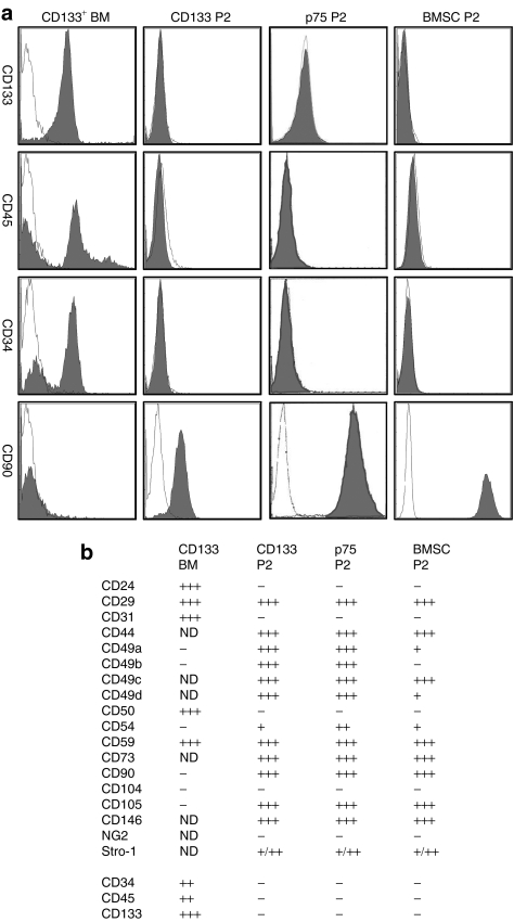
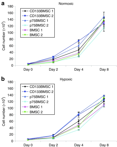
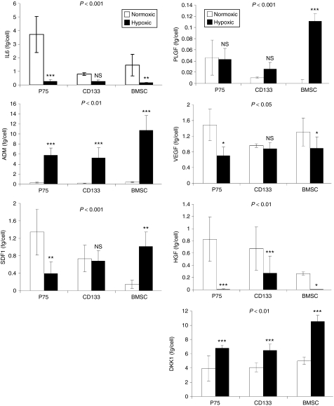
The reparative properties of bone marrow stromal cells (BMSCs) have been attributed in part to the paracrine action of secreted factors. We isolated typical human BMSCs by plastic adherence and compared them with BMSC sub-populations isolated by magnetic-activated cell sorting against CD133 (CD133-derived BMSCs, CD133BMSCs) or CD271 [p75 low-affinity nerve growth factor receptor (p75LNGFR), p75BMSCs]. Microarray assays of expressed genes, and enzyme-linked immunosorbent assays (ELISAs) of selected growth factors and cytokines secreted under normoxic and hypoxic conditions demonstrated that the three transit-amplifying progenitor cell populations were distinct from one another. CD133BMSC-conditioned medium (CdM) was superior to p75BMSC CdM in protecting neural progenitor cells against cell death during growth factor/nutrient withdrawal. Intracardiac (arterial) administration of concentrated CD133BMSC CdM provided neuroprotection and significantly reduced cortical infarct volumes in mice following cerebral ischemia. In support of the paracrine hypothesis for BMSC action, intra-arterial infusion of CD133BMSC CdM provided significantly greater protection against stroke compared with the effects of CD133BMSC (cell) administration. CdM from CD133BMSCs also provided superior protection against stroke compared with that conferred by CdM from p75BMSCs or typically isolated BMSCs. CD133 identifies a sub-population of nonhematopoietic stem/progenitor cells from adult human bone marrow, and CD133BMSC CdM may provide neuroprotection for patients with stroke.


Figures

<b>Figure 1</b>
Figure 1
Cell surface epitopes. (a) Illustrating changes in cell surface epitope expression that occur in freshly isolated CD133+ cells from human bone marrow (CD133 BM) before and after they adhere to generate CD133BMSCs. Over time, the CD133 BM cells lose the expression of CD133 and acquire the expression of typical adherent BMSC markers such as CD90 (Thy 1) and CD105 (endoglin). Passage 2 CD133BMSCs (CD133 P2), p75BMSCs (p75 P2), and typical BMSCs (BMSC P2) are negative for CD34 and the pan-hematopoietic marker CD45. Antibody staining is shown in dark fill and isotype staining is shown in white fill. (b) Summary for all epitopes tested. Two donors were stained for each cell type. +, 1–25% cells positive; ++, 25–50% cells positive; +++, 50–100% cells positive; BMSC, bone marrow stromal cell; ND, not determined.
<b>Figure 2</b>
Figure 2
Multipotent differentiation of CD133BMSCs and p75BMSCs. (a–c) Phase contrast photomicrographs of cultured CD133BMSCs, p75BMSCs, and typical BMSCs. Bar = 50 µm. (d–f) Differentiation of CD133BMSCs into osteoblasts, adipocytes, and chondrocytes, respectively. Bar = 1 mm. (g–i) Differentiation of p75BMSCs into osteoblasts and adipocytes, but not chondrocytes. The p75BMSCs form a micromass pellet (i) but do not possess typical chondrocyte morphology. g, bar = 1 mm; h,i, bar = 62.5 µm. Calcification is stained by Alizarin Red S (d,g). Lipid is stained by Oil Red O (e,h). Sulfated proteoglycans are stained by toluidine blue sodium borate (f,i). BMSC, bone marrow stromal cell.
<b>Figure 3</b>
Figure 3
Growth of CD133BMSCs, p75BMSCs, and BMSCs under normoxic and hypoxic conditions. BMSCs isolated by typical plastic adherence as well as those isolated by magnetic-activated cell sorting against CD133 (CD133BMSC) or p75LNGFR (p75BMSC) grow equally well under (a) normoxic and (b) hypoxic (1% oxygen) conditions. Cell growth data are shown for two donors for each cell type over 8 days. Cells from all of the donors were plated at 100 cells/cm2 and allowed to grow for 2 days in a normoxic incubator prior to moving half of the plates to a hypoxic incubator to begin the assay (day 0). There was no significant difference in cell growth rates for either condition (two-way analysis of variance). BMSC, bone marrow stromal cell; p75LNGFR, p75 low-affinity nerve growth factor receptor.
<b>Figure 4</b>
Figure 4
Microarray assays of expressed genes. Top: hierarchical clustering for gene expression for p75LNGFR+ and CD133+ cells freshly isolated from human bone marrow mononuclear cells and passage 2 (P2) BMSCs, CD133BMSCs, and p75BMSCs cultured in complete culture medium. The overall transcriptional profiles of the p75BMSC and CD133BMSC sub-populations are more similar to each other than to the profile for typical BMSCs. Middle: heat map depicting gene expression. Note, nine major patterns with patterns 6, 9, and 4 in particular demonstrating differentially upregulated genes for p75BMSCs, CD133BMSCs, and BMSCs, respectively (see also Supplementary Table S2). Bottom: the numbers of transcripts contained in each pattern are listed with the numbers of uniquely expressed genes for each pattern shown in parentheses. BMSC, bone marrow stromal cell; p75LNGFR, p75 low-affinity nerve growth factor receptor.
<b>Figure 5</b>
Figure 5
Growth factor/cytokine secretion under normoxic and hypoxic conditions. Levels of selected secreted proteins/peptides for cells grown to 90% confluence. N = 3 donors in each case. Enzyme-linked immunosorbent assay (ELISA) data were normalized for cell number. Statistical significance (P values) pictured at the tops of the graphs indicate the degree to which the cell populations responded differently from each other when exposed to hypoxia (repeated measures analysis of variance). The asterisks denote differences in protein/peptide secretion levels for a particular cell type in normoxic versus hypoxic conditions. ADM, adrenomedullin; BMSC, bone marrow stromal cell; DKK1, Dickkopf-1; HGF, hepatocyte growth factor; IL6, interleukin 6; NS, not significant; PLGF, placental growth factor; SDF1, stromal-derived factor 1; VEGF, vascular endothelial growth factor. *P ≤ 0.05, **P ≤ 0.01, ***P ≤ 0.001. All ELISA measurements were performed in triplicate.
<b>Figure 6</b>
Figure 6
Protection of neural progenitor cells (NPCs) from cell death induced by growth factor withdrawal. (a) Neural stem cells isolated as neurospheres from day 4 postnatal GFP mice. (b) Differentiation of NPCs into immature neurons (βIII-tubulin-positive; ALEXA 594, red). (c) Differentiation of NPCs into astrocytes (GFAP+ ALEXA 594, red). (d) Percent survival for NPCs after plating onto laminin/poly-D-lysine coated cell ware and growth factor withdrawal for 48 hours during incubation in serum-free alpha minimum essential medium (SFM), or conditioned medium (CdM) from p75BMSCs (p75, n = 3 donors), CD133BMSCs (CD133, n = 3 donors), or typical BMSCs (BMSC, n = 2 donors). MTS measurements were performed in triplicate. **P ≤ 0.01 compared with SFM, P ≤ 0.01 compared with p75 CdM. BMSC, bone marrow stromal cell; GFAP, glial fibrillary acidic protein; GFP, green fluorescent protein; MTS, 3-(4,5-dimethylthiazol-2-yl)-5-(3-carboxymethoxyphenyl)-2-(4-sulfophenyl)-2H-tetrazolium.
<b>Figure 7</b>
Figure 7
Protection against cerebral ischemia. (a) Representative 2,3,5-triphenyltetrazolium chloride (TTC) stains to indicate viable cortical tissue in sham-operated animal, and 1 or 3 days after middle cerebral artery ligation (MCAL). In sham surgery, the needle is passed under the middle cerebral artery, but the suture is not tied. Bar = 1 mm. (b) Representative cresyl violet stains of brain sections from immunodeficient mice that underwent middle cerebral artery ligation surgery and treatment 24 hours later with PBS, 2 million human CD133BMSCs (cells), or concentrated CdM from p75BMSCs, CD133BMSCs, typical BMSCs (BMSC), or human dermal fibroblasts. PBS vehicle, CD133BMSCs, or CdM from the different cell types was injected into the left ventricle of the heart (intra-arterial, 100 µl). Animals were euthanized 48 hours following treatment for analysis. Bar = 1 mm. (c) Quantification of the cortical infarct volumes for PBS-treated (PBS, n = 5), p75BMSC CdM–treated (p75, n = 5), CD133BMSC-treated (cells, n = 6), CD133BMSC CdM–treated (CD133, n = 5), BMSC CdM–treated (BMSC, n = 5), and fibroblast CdM–treated mice (Fibro, n = 6). *P < 0.05 compared with PBS, **P < 0.01 compared with PBS. Statistics were determined by analysis of variance with Bonferroni post hoc testing. BMSC, bone marrow stromal cell; CdM, conditioned medium; PBS, phosphate-buffered saline.

References

    1. Till JE., and , McCulloch EA. A direct measurement of the radiation sensitivity of normal mouse bone marrow cells. Radiat Res. 1961;14:213–222. - PubMed
    1. Till JE, McCulloch EA, Siminovitch L. A stochastic model of stem cell proliferation, based on the growth of spleen colony-forming cells. Proc Natl Acad Sci U S A. 1964;51:29–36. - PMC - PubMed
    1. Morrison SJ, Uchida N., and , Weissman IL. The biology of hematopoietic stem cells. Annu Rev Cell Dev Biol. 1995;11:35–71. - PubMed
    1. Akashi K, Traver D, Miyamoto T., and , Weissman IL. A clonogenic common myeloid progenitor that gives rise to all myeloid lineages. Nature. 2000;404:193–197. - PubMed
    1. Kondo M, Weissman IL., and , Akashi K. Identification of clonogenic common lymphoid progenitors in mouse bone marrow. Cell. 1997;91:661–672. - PubMed

Publication types

MeSH terms