

doi: 10.1038/nchembio0909-602.
Working towards an exegesis for lipids in biology
Affiliations
- PMID: 19690530
- PMCID: PMC3785062
- DOI: 10.1038/nchembio0909-602
Item in Clipboard
Working towards an exegesis for lipids in biology
Nat Chem Biol.
2009 Sep.
Abstract
As a field, lipidomics is in its infancy, yet it has already begun to influence lipid biochemistry in myriad ways. As with other omic technologies, the field is driven by advances in analytical chemistry, particularly by mass spectrometry. At the heart of a renaissance in lipid biochemistry, systems biology is being used to define the cellular lipome, build a comprehensive picture of metabolic interconnections, discover new molecular species and determine how lipids modulate biological functions.
Figures
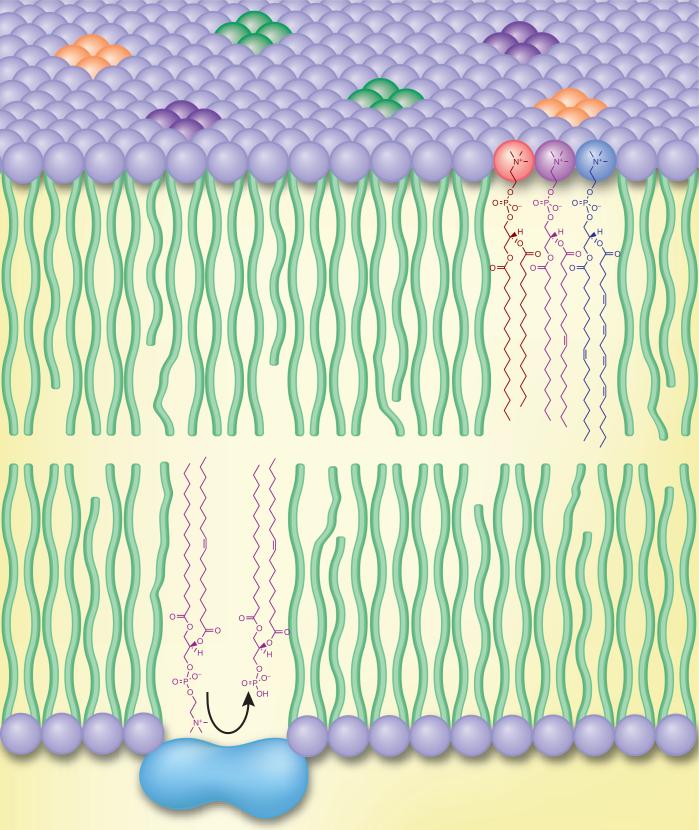
Biological membranes are often depicted as a sea of homogenous lipid in which membrane proteins reside and cytosolic proteins translocate. In reality, cellular membranes are composed of chemically diverse lipid species that regulate essential biophysical, metabolic and signaling processes.
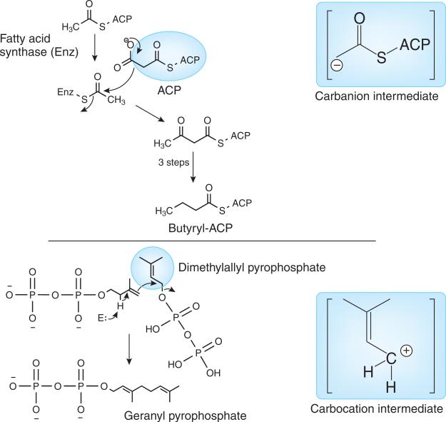
Known initial biochemical steps responsible for the synthesis of all lipids. Synthesis occurs via either a carbanion intermediate (fatty acyls and polyketides, hence mammalian and plant glycerolipids, sphingolipids, saccharolipids and glycerophospholipids) or a carbocation intermediate (prenols, steroids and archaeal glycerolipids, sphingolipids and glycerophospholipids), according to Fahy et al..

Brain PC imaged by MALDI mass spectrometry. Mass spectral imaging of a 10 μm mouse brain slice (mid-sagittal section) for a docosahexaenoic acid–containing phosphatidylcholine (18:0/22:6-PC) as a positive ion at m/z 834.6 using MALDI mass spectrometry. The highest ion intensity (brightest shade) was observed in the cerebellar gray matter (see ref. for methods). c.p.s., counts per second.
References
Publication types
MeSH terms
Substances
Grants and funding
LinkOut - more resources
Full Text Sources
