

Clinical anatomy of the quadriceps femoris and extensor apparatus of the knee
- PMID: 19690926
- PMCID: PMC2772911
- DOI: 10.1007/s11999-009-1052-y
Clinical anatomy of the quadriceps femoris and extensor apparatus of the knee
Abstract
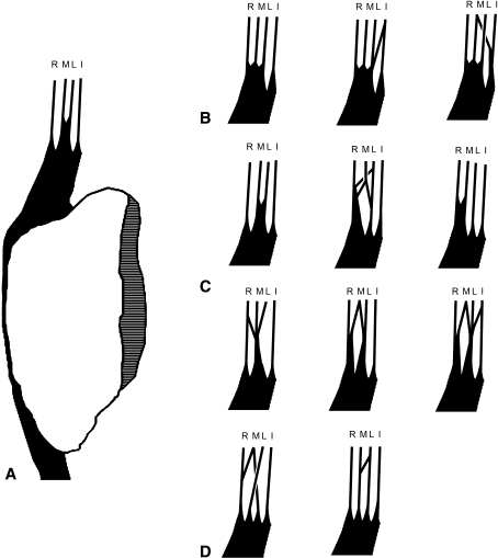
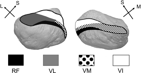
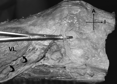
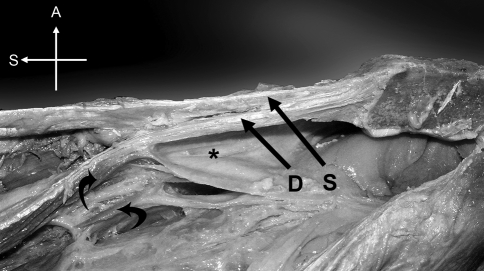
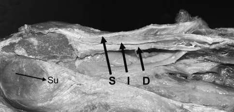
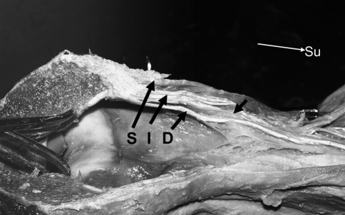
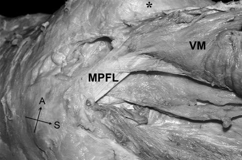
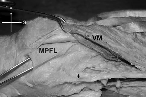
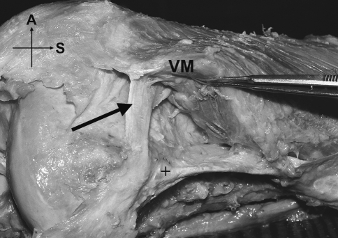
Most descriptions of the extensor mechanism of the knee do not take into account its complexity and variability. The quadriceps femoris insertion into the patella is said to be through a common tendon with a three-layered arrangement: rectus femoris (RF) most superficially, vastus medialis (VM) and lateralis (VL) in the intermediate layer, and vastus intermedius (VI) most deeply. We dissected 20 limbs from 17 cadavers to provide a more detailed description of the anterior components of the knee: the tendon, the patellar retinacula, and the patellofemoral ligaments. Only three of the 20 specimens exhibited the typically described quadriceps pattern. The remainder had bilaminar and even more complex trilaminar and tetralaminar fiber arrangements. We found an oblique head of the vastus lateralis (VLO), separated from the longitudinal head by a layer of fat or fascia, in 60% of the specimens. However, we found no distinct oblique head of the vastus medialis (VMO) in any specimen. The medial patellofemoral ligament (MPFL) was more common than the lateral (LPFL), supporting its suggested role as the principal passive medial stabilizer of the patella. Because the quadriceps muscle group plays a direct role in patellofemoral joint function, investigation into the clinical applications of its highly variable anatomy may be worthwhile with respect to joint dysfunction and failures of TKAs.
Figures
References
-
- {'text': '', 'ref_index': 1, 'ids': [{'type': 'DOI', 'value': '10.1097/JSA.0b013e318053eb74', 'is_inner': False, 'url': 'https://doi.org/10.1097/jsa.0b013e318053eb74'}, {'type': 'PubMed', 'value': '17505317', 'is_inner': True, 'url': 'https://pubmed.ncbi.nlm.nih.gov/17505317/'}]}
- Amis AA. Current concepts on anatomy and biomechanics of patellar stability. Sports Med Arthrosc. 2007;15:48–56. - PubMed
-
- {'text': '', 'ref_index': 1, 'ids': [{'type': 'DOI', 'value': '10.1016/S0968-0160(03)00006-1', 'is_inner': False, 'url': 'https://doi.org/10.1016/s0968-0160(03)00006-1'}, {'type': 'PubMed', 'value': '12893142', 'is_inner': True, 'url': 'https://pubmed.ncbi.nlm.nih.gov/12893142/'}]}
- Amis AA, Firer P, Mountney J, Senavongse W, Thomas NP. Anatomy and biomechanics of the medial patellofemoral ligament. Knee. 2003;10:215–220. - PubMed
-
- {'text': '', 'ref_index': 1, 'ids': [{'type': 'DOI', 'value': '10.1007/s00167-005-0680-3', 'is_inner': False, 'url': 'https://doi.org/10.1007/s00167-005-0680-3'}, {'type': 'PubMed', 'value': '16283173', 'is_inner': True, 'url': 'https://pubmed.ncbi.nlm.nih.gov/16283173/'}]}
- Andrikoula S, Tokis A, Vasiliadis HS, Georgoulis A. The extensor mechanism of the knee joint: an anatomical study. Knee Surg Sports Traumatol Arthrosc. 2006;14:214–220. - PubMed
-
- {'text': '', 'ref_index': 1, 'ids': [{'type': 'PubMed', 'value': '11046166', 'is_inner': True, 'url': 'https://pubmed.ncbi.nlm.nih.gov/11046166/'}]}
- Bencardino JT, Rosenberg ZS, Brown RR, Hassankhani A, Lustrin ES, Beltran J. Traumatic musculotendinous injuries of the knee: diagnosis with MR imaging. Radiographics. 2000;20:S103–S120. - PubMed
-
- {'text': '', 'ref_index': 1, 'ids': [{'type': 'DOI', 'value': '10.1590/S1413-78522004000200005', 'is_inner': False, 'url': 'https://doi.org/10.1590/s1413-78522004000200005'}]}
- Bevilaqua-Grossi D, Monteiro-Pedro V, Bérzin F. Functional analysis of the patellar stabilizers. Acta Ortop Bras. 2004;12:99–104.
MeSH terms
LinkOut - more resources
Full Text Sources
