

Tibialis posterior in health and disease: a review of structure and function with specific reference to electromyographic studies
- PMID: 19691828
- PMCID: PMC2739849
- DOI: 10.1186/1757-1146-2-24
Tibialis posterior in health and disease: a review of structure and function with specific reference to electromyographic studies
Abstract
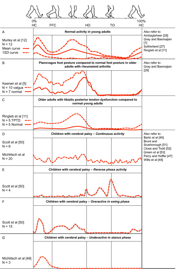
Tibialis posterior has a vital role during gait as the primary dynamic stabiliser of the medial longitudinal arch; however, the muscle and tendon are prone to dysfunction with several conditions. We present an overview of tibialis posterior muscle and tendon anatomy with images from cadaveric work on fresh frozen limbs and a review of current evidence that define normal and abnormal tibialis posterior muscle activation during gait. A video is available that demonstrates ultrasound guided intra-muscular insertion techniques for tibialis posterior electromyography.Current electromyography literature indicates tibialis posterior intensity and timing during walking is variable in healthy adults and has a disease-specific activation profile among different pathologies. Flat-arched foot posture and tibialis posterior tendon dysfunction are associated with greater tibialis posterior muscle activity during stance phase, compared to normal or healthy participants, respectively. Cerebral palsy is associated with four potentially abnormal profiles during the entire gait cycle; however it is unclear how these profiles are defined as these studies lack control groups that characterise electromyographic activity from developmentally normal children. Intervention studies show antipronation taping to significantly decrease tibialis posterior muscle activation during walking compared to barefoot, although this research is based on only four participants. However, other interventions such as foot orthoses and footwear do not appear to systematically effect muscle activation during walking or running, respectively. This review highlights deficits in current evidence and provides suggestions for the future research agenda.
Figures
References
-
- Basmajian J, Stecko G. The role of muscles in support of the arch of the foot. J Bone Joint Surg Am. 1963;45:1184–1190. - PubMed
-
- Kaye RA, Jahss MH. Tibialis posterior: a review of anatomy and biomechanics in relation to support of the medial longitudinal arch. Foot Ankle. 1991;11:244. - PubMed
-
- Myerson M. Adult acquired flatfoot deformity: treatment of dysfunction of the posterior tibial tendon. Instr Course Lect. 1997;46:393–405. - PubMed
-
- Myerson M, Solomon G, Shereff M. Posterior tibial tendon dysfunction: its association with seronegative inflammatory disease. Foot Ankle. 1989;9:219–225. - PubMed
-
- Keenan M, Peabody T, Gronley J, Perry J. Valgus deformities of the feet and characteristics of gait in patients who have rheumatoid arthritis. J Bone Joint Surg Am. 1991;73:237–247. - PubMed
Grants and funding
LinkOut - more resources
Full Text Sources
Other Literature Sources
Medical
Molecular Biology Databases
