. 2009 Oct 16;284(42):28823-31.
doi: 10.1074/jbc.M109.039123. Epub 2009 Aug 18.

Dbf4-Cdc7 phosphorylation of Mcm2 is required for cell growth

Affiliations

Dbf4-Cdc7 phosphorylation of Mcm2 is required for cell growth

Irina Bruck et al. J Biol Chem. .

Abstract

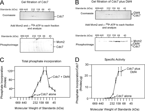
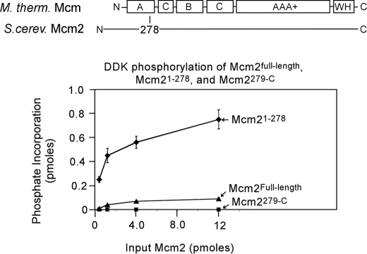
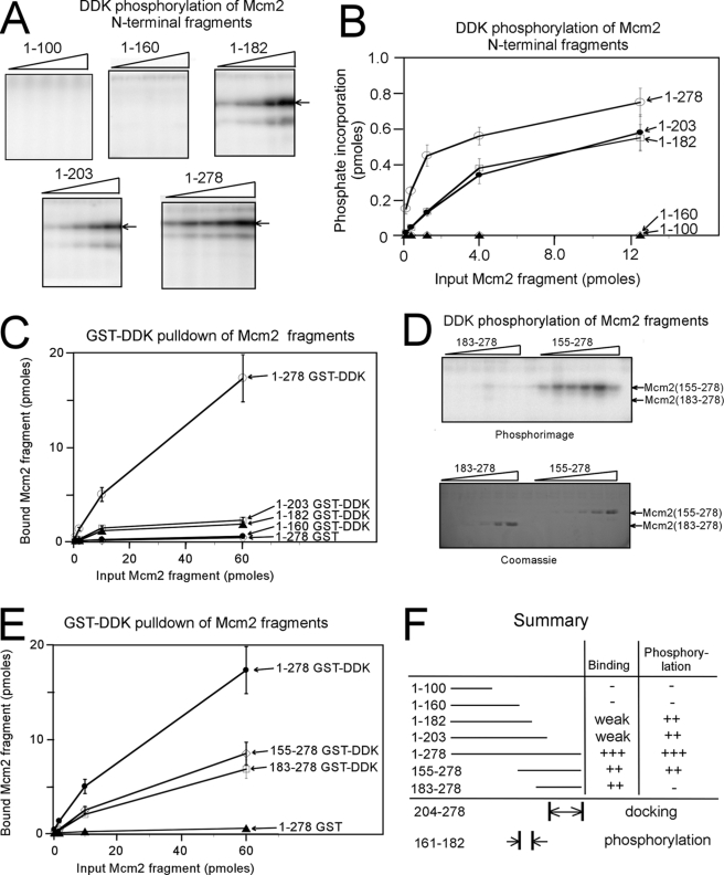
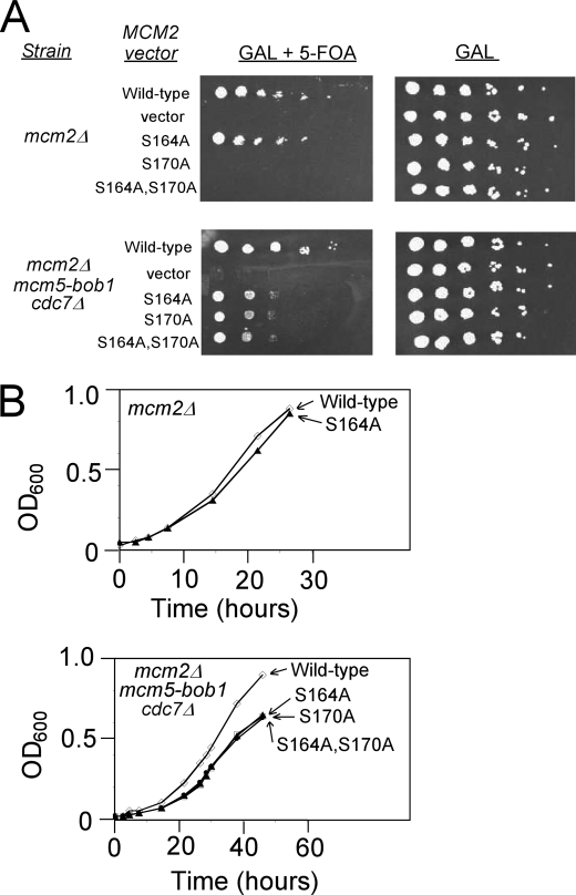
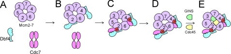
The Dbf4-Cdc7 kinase (DDK) is required for the activation of the origins of replication, and DDK phosphorylates Mcm2 in vitro. We find that budding yeast Cdc7 alone exists in solution as a weakly active multimer. Dbf4 forms a likely heterodimer with Cdc7, and this species phosphorylates Mcm2 with substantially higher specific activity. Dbf4 alone binds tightly to Mcm2, whereas Cdc7 alone binds weakly to Mcm2, suggesting that Dbf4 recruits Cdc7 to phosphorylate Mcm2. DDK phosphorylates two serine residues of Mcm2 near the N terminus of the protein, Ser-164 and Ser-170. Expression of mcm2-S170A is lethal to yeast cells that lack endogenous MCM2 (mcm2Delta); however, this lethality is rescued in cells harboring the DDK bypass mutant mcm5-bob1. We conclude that DDK phosphorylation of Mcm2 is required for cell growth.


Figures

FIGURE 1.
FIGURE 1.
Low molecular weight form of Dbf4-Cdc7 exhibits high specific activity for Mcm2. A, 200 μg of Cdc7 alone was subjected to Superose 6 size exclusion chromatography as described under “Experimental Procedures.” Each fraction was then subjected to SDS-PAGE analysis followed by Coomassie staining (top gel). The relative positions of molecular weight standards analyzed under identical conditions are shown above the gel. Each column fraction was incubated with 500 ng of full-length Mcm2 and [γ-32P]ATP for 30 min at 30 °C, and the reactions were analyzed by SDS-PAGE followed by phosphorimaging (bottom gel). The migration distances of Mcm2 and Cdc7, shown at the right of the gel, were determined by analyzing purified standards in the same gel. B, 200 μg of Cdc7 was incubated with 50 μg of Dbf4 for 30 min at 15 °C, and the mixture was then subjected to Superose 6 size exclusion chromatography as in A. The fractions were analyzed for protein abundance (top gel) and Mcm2 phosphorylation (bottom gel) as in A. C, quantitation of phosphate incorporation from the phosphorimages of A and B, and similar experiments are plotted as a function of size exclusion fraction (mean ± S.E., n = 3). The relative elution of molecular weight standards is shown at the bottom of the graph. D, amount of Cdc7 present in each reaction of C was quantified by densitometry of the Coomassie images. The phosphate incorporation into Mcm2 per μg of Cdc7 was calculated and plotted as function of gel filtration fraction.
FIGURE 2.
FIGURE 2.
Dbf4 or DDK, but not Cdc7, binds tightly to Mcm2 and Mcm2-7. A, Mcm2 or Mcm2-7 bearing a site for protein kinase A phosphorylation (PKA site on Mcm3) was radiolabeled with protein kinase A as described under “Experimental Procedures.” 50 pmol of GST-DDK, GST-Dbf4, GST-Cdc7, or GST alone was incubated with varying concentrations of radiolabeled full-length Mcm2 (left) or Mcm2-7 complex (right) for 1 h at room temperature. For Mcm2, the total volume for each reaction was 100 μl, and the quantity of Mcm2 added was 66, 20, or 6.6 pmol. For Mcm2-7, the total volume for each reaction was 100 μl, and the quantity of Mcm2-7 complex added was 20, 6.6, or 2 pmol. After mixing, 40 μl of glutathione-Sepharose beads was added to each reaction, and the centrifuged pellet was washed and analyzed by SDS-PAGE followed by phosphorimaging. Input for the GST-pulldown was analyzed in the same gel to quantify the amount of protein bound. Results from experiments similar to those shown in A were quantified and plotted for Mcm2 in B and for Mcm2-7 in C. The mean ± S.E. is shown for each input amount.
FIGURE 3.
FIGURE 3.
DDK phosphorylates the N-terminal region of Mcm2. A structure-based sequence alignment with Sulfolobus solfataricus Mcm was used to divide Saccharomyces cerevisiae Mcm2 into two segments, an N-terminal fragment encompassing the nonconserved N-terminal region and most of domain A (Mcm2-(1–278)) and a C-terminal fragment encompassing the rest of the protein (Mcm2-(279-C)). S. cerevisiae Mcm2Full-length, Mcm2-(1–278), or Mcm2-(279-C) was incubated with 50 ng of DDK and [γ-32P]ATP in a volume of 10 μl for 30 min at 30 °C, and the reactions were then analyzed by SDS-PAGE followed by phosphorimaging. The results were quantified, and the fraction of phosphate incorporation is plotted as a function of input in picomoles.
FIGURE 4.
FIGURE 4.
Region 161–182 of Mcm2 is critical for DDK phosphorylation. A, N-terminal fragments of Mcm2 of varying amounts were incubated with 50 ng of DDK and [γ-32P]ATP in a volume of 10 μl for 30 min at 30 °C. The amount of Mcm2 fragment added was 13, 4, 1.3, 0.4, and 0.13 pmol. The reactions were then analyzed by SDS-PAGE followed by phosphorimaging. B, results from experiments similar to A were quantified, and the fraction of phosphate incorporation is plotted as a function of input in picomoles. The data are mean ± S.E. C, N-terminal fragments of Mcm2 with a site for protein kinase A phosphorylation was radiolabeled with [γ-32P]ATP and PKA as described under “Experimental Procedures.” 50 pmol of GST-DDK was incubated with varying amounts of radiolabeled Mcm2 fragments for 1 h at room temperature in a total volume of 100 μl. After mixing, the reactions were analyzed as described under “Experimental Procedures.” D, fragments 155–278 and 183–278 of Mcm2 of were tested for DDK phosphorylation as described in A. The amount of Mcm2 fragment added was 130, 40, 13, 4, 1.3, 0.4, and 0.13 pmol. The reactions were then analyzed by SDS-PAGE followed by phosphorimaging (top gel) or Coomassie staining (bottom gel). E, fragments of Mcm2 were analyzed by a GST-pulldown assay with GST-DDK as described in C. F, summary of data from A to E.
FIGURE 5.
FIGURE 5.
DDK phosphorylates Ser-164 and Ser-170 of Mcm2. A, residues 161–181 of S. cerevisiae Mcm2 are shown, and Ser-164 and Ser-170 were targeted for site-directed mutagenesis. B, mutants of Mcm2-(1–278) of varying amounts were incubated with 50 ng of DDK and [γ-32P]ATP in a volume of 10 μl for 30 min at 30 °C. The amount of Mcm2-(1–278) added was 12 or 4 pmol (left image) or 1.2 or 0.4 pmol (right image). The reactions were then analyzed by SDS-PAGE followed by phosphorimaging. C, results from experiments similar to B were quantified, and the fraction of phosphate incorporation is plotted as a function of input in picomoles. The data are mean ± S.E. D, mutants of Mcm2-(1–278) with a site for protein kinase A phosphorylation were radiolabeled with [γ-32P]ATP and protein kinase A (PKA) as described under “Experimental Procedures.” 50 pmol of GST-Dbf4 was incubated with varying amounts of radiolabeled Mcm2 fragments for 1 h at room temperature in a total volume of 100 μl. After mixing, the reactions were analyzed as described under “Experimental Procedures.” E, sequence alignment of budding yeast Mcm2 and budding yeast Mcm4. Serine and threonine residues are in boldface, and acidic residues are underlined. The aligned regions of these two proteins contain phosphoacceptor sites for DDK. F, alignment of budding yeast Mcm2 with Mcm2 from higher eukaryotes. DDK phosphoacceptor sites of human Mcm2 do not map to this region (10, 19, 20).
FIGURE 6.
FIGURE 6.
DDK phosphorylation of Mcm2 Ser-170 is required for budding yeast growth. A, budding yeast cells deleted for mcm2 (mcm2Δ, top plates) or deleted for mcm2 and cdc7 and bearing the mcm5-bob1 mutation (mcm2Δ, mcm5-bob1, and cdc7Δ, bottom plates) were used. The cells harbor wild-type MCM2 on a plasmid with a URA3 selectable marker and mutant mcm2 on a LEU2 plasmid with expression controlled by a galactose-inducible promoter. In the presence of galactose and 5-FOA, only mcm2 under the control of the galactose-inducible promoter is expressed (left plates). In the presence of galactose (right plates), wild-type MCM2 is expressed as well. Cells were plated in serial 10-fold dilutions. B, colonies from A were grown in liquid media, and the rate of growth was measured as function of time. The top graph is for mcm2Δ cells, and the bottom graph is for mcm2Δ,mcm5-bob1,cdc7Δ cells.
FIGURE 7.
FIGURE 7.
Model for DDK phosphorylation of Mcm2-7. A, Cdc7 is weakly active as a multimer on its own. B, Dbf4 binds tightly to residues 203–378 of Mcm2. Dbf4 likely binds to the homologous region of Mcm4 as well. C, Dbf4 recruits a monomer of Cdc7 to Mcm2 to activate the kinase, phosphorylating Ser-170 of Mcm2. The homologous region of Mcm4 is likely phosphorylated as well (26). D, phosphorylation of Ser-170 of Mcm2 induces a conformational change in the Mcm2-7 complex that is mimicked by the mcm5-bob1 mutation. E, conformational change in Mcm2-7 allows for the proper positioning of Cdc45 and GINS, and the Mcm2-7-Cdc45-GINS complex is activated to unwind DNA. DDK may also phosphorylate other Mcm proteins (data not shown).

References

    1. Bousset K., Diffley J. F. (1998) Genes Dev. 12, 480–490 - PMC - PubMed
    1. Donaldson A. D., Fangman W. L., Brewer B. J. (1998) Genes Dev. 12, 491–501 - PMC - PubMed
    1. Tanaka S., Tak Y. S., Araki H. (2007) Cell Div. 2, 16. - PMC - PubMed
    1. Tanaka S., Umemori T., Hirai K., Muramatsu S., Kamimura Y., Araki H. (2007) Nature 445, 328–332 - PubMed
    1. Zegerman P., Diffley J. F. (2007) Nature 445, 281–285 - PubMed

Publication types

MeSH terms

Substances