. 2009 Nov;104(6):1141-9.
doi: 10.1093/aob/mcp199. Epub 2009 Aug 18.

Elaiophores in Gomesa bifolia (Sims) M.W. Chase & N.H. Williams (Oncidiinae: Cymbidieae: Orchidaceae): structure and oil secretion

Affiliations

Elaiophores in Gomesa bifolia (Sims) M.W. Chase & N.H. Williams (Oncidiinae: Cymbidieae: Orchidaceae): structure and oil secretion

Sandra S Aliscioni et al. Ann Bot. 2009 Nov.

Abstract

Background and aims: Oils are an unusual floral reward in Orchidaceae, being produced by specialized glands called elaiophores. Such glands have been described in subtribe Oncidiinae for a few species. The aims of the present study were to identify the presence of elaiophores in Gomesa bifolia, to study their structure and to understand how the oil is secreted. Additionally, elaiophores of G. bifolia were compared with those of related taxa within the Oncidiinae.

Methods: Elaiophores were identified using Sudan III. Their structure was examined by using light, scanning electron and transmission electron microscopy.

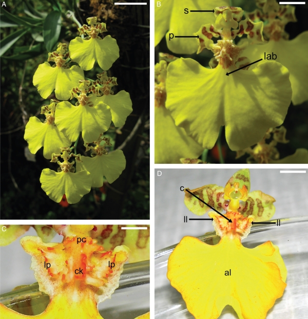
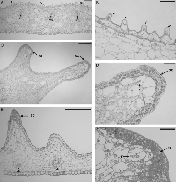
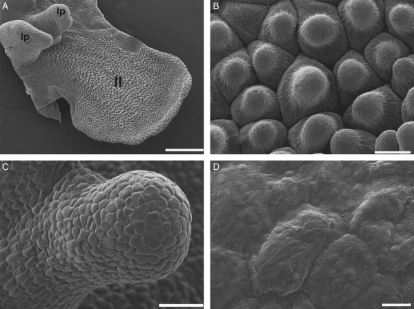
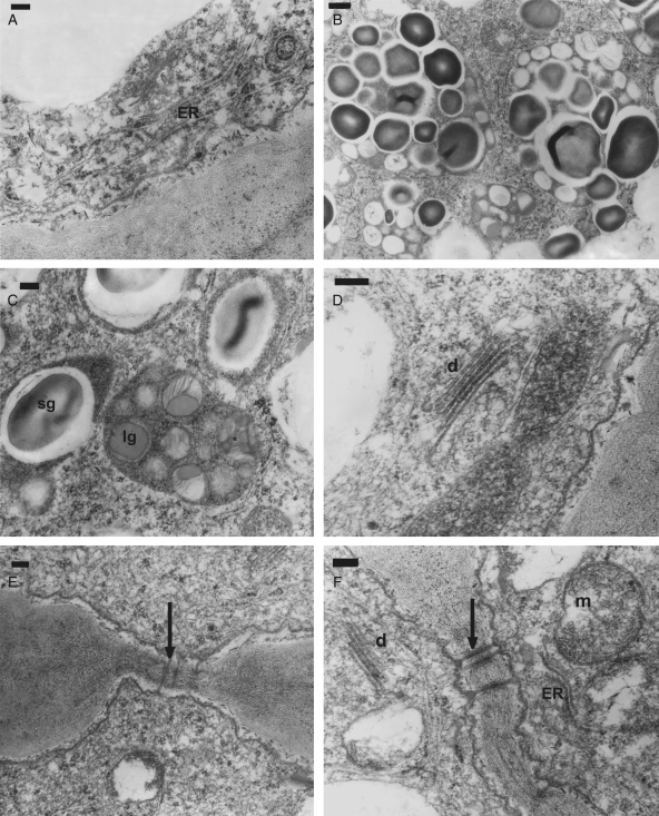
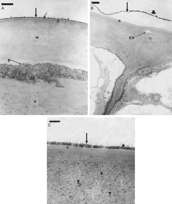
Key results: Secretion of oils was from the tips of callus protrusions. The secretory cells each had a large, centrally located nucleus, highly dense cytoplasm, abundant plastids containing lipid globules associated with starch grains, numerous mitochondria, an extensive system of rough and smooth endoplasmatic reticulum, and electron-dense dictyosomes. The outer tangential walls were thick, with a loose cellulose matrix and a few, sparsely distributed inconspicuous cavities. Electron-dense structures were observed in the cell wall and formed a lipid layer that covered the cuticle of the epidermal cells. The cuticle as viewed under the scanning electron microscope was irregularly rugose.

Conclusions: The elaiophores of G. bifolia are of the epithelial type. The general structure of the secretory cells resembles that described for other species of Oncidiinae, but some unique features were encountered for this species. The oil appears to pass through the outer tangential wall and the cuticle, covering the latter without forming cuticular blisters.


Figures

Fig. 1.
Fig. 1.
General view of Gomesa bifolia. (A) Inflorescence. (B) Flower showing sepal (s), petal (p) and labellum (lab). (C, D) Flower stained with Sudan III: (C) detail of the callus showing central keel (ck), proximal crest (pc) and lateral protuberances (lp); (D) labellum showing apical lobe (al), lateral lobes (ll) and callus (c). Scale bars: (A) = 1·5 cm, (B) = 0·5 cm, (C) = 0·2 cm, (D) = 0·6 cm.
Fig. 2.
Fig. 2.
Light micrographs of transverse sections of the lateral lobe and callus of Gomesa bifolia. (A, B) Lateral lobe: (A) section with vascular bundles (vb) and papillose cells (arrows); (B) detail of the papillose cells (arrows). (C, D) Lateral protuberances of the callus: (C) section showing two protrusions with secretory cells on the tips (sc); (D) detail of the tip of the protuberance showing secretory cells (sc) and raphides in the parenchyma (r). (E, F) Proximal crest of the callus: (E) section showing vascular bundles (vb) and secretory cells on the tip of a protrusion (sc); (F) detail of the tip of protrusion showing secretory cells (sc) and raphides in the parenchyma (r). Scale bars: (A) = 120 µm; (B) = 60 µm; (C) = 280 µm; (D, F) = 70 µm; (E) = 250 µm.
Fig. 3.
Fig. 3.
Scanning electron micrographs of the lateral lobe and callus of Gomesa bifolia. (A) Surface of a lateral lobe (ll) and two lateral protuberances of the callus (lp). (B) Detail of the papillae in the lateral lobe. (C, D) Lateral protuberance of the callus: (C) general view of a lateral protuberance; (D) epidermal cells on the tip. Scale bars: (A) = 500 µm, (B) = 20 µm, (C) = 100 µm, (D) = 10 µm.
Fig. 4.
Fig. 4.
TEM sections through an elaiophore of Gomesa bifolia. (A–F) Section of secretory cells on the tip of the callus protrusions and their complement of organelles. (A) Detail of cytoplasm with abundant endoplasmic reticulum (ER). (B, C) Detail of plastids with starch grains (sg) and lipid globules (lg). (D) Detail of cytoplasm with dictyosomes (d). (E) Detail of the connections (arrow) between epidermal and subepidermal cells. (F) Detail of the connections (arrow) between radial walls of the secretory epidermal cells; cytoplasm showing mitochondria (m), dictyosomes (d) and endoplasmic reticulum (ER). Scale bars: (A, C–F) = 0·2 µm; (B) = 0·5 µm.
Fig. 5.
Fig. 5.
TEM sections through an elaiophore of Gomesa bifolia. (A–C) Section of the outer tangencial wall of the secretory cells on the tips of the callus protrusions. (A) Detail of outer tangential wall covered by an electron-dense layer (arrow); cuticle (c), cell wall (w), plasmalemma (p), cytoplasm (cy) and vacuole (v) are indicated. (B) Outer tangential wall showing a small cavity (cv) and detached electron-dense layer (arrow). (C) Detail of the outer tangential wall showing the cuticle (c), electron-dense corpuscles (arrowheads) and lipid layer (arrow). Scale bars: (A, B) = 1 µm; (C) = 0·2 µm.

References

    1. Ackerman JD. Pollination of tropical and temperate orchids. In: Tan KW, editor. Proceedings of the 11th World Orchid Conference; American Orchid Society; Miami, FL. 1984. pp. 98–101.
    1. Buchmann SL. The ecology of oil flowers and their bees. Annual Review of Ecology and Systematics. 1987;18:343–369.
    1. Chase MW, Palmer JD. Floral morphology and chromosome number in subtribe Oncidiinae (Orchidaceae): evolutionary insights from a phylogenetic analysis of chloroplast DNA restriction site variation. In: Soltis PS, Soltis DE, Doyle JJ, editors. Molecular systematics of plants. New York: Chapman and Hall; 1992. pp. 324–339.
    1. Chase MW, Cameron KN, Barrett RL, Freudenstein JV. DNA data and Orchidaceae systematics: a new phylogenetic classification. In: Dixon KM, Kell SP, Barrett RL, Cribb PJ, editors. Orchid conservation. Kota Kinabalu, Sabah: Natural History Publications; 2003. pp. 69–89.
    1. Chase MW, Williams NH, de Faria AD, Neubig KM, Amaral MCE, Whitten WM. Floral convergence in Oncidiinae (Cymbidieae; Orchidaceae): an expanded concept of Gomesa and a new genus Nohawilliamsi. Annals of Botany. 2009;104:387–402. - PMC - PubMed

MeSH terms