Review
. 2009 Sep 1;122(Pt 17):3037-49.
doi: 10.1242/jcs.052704.

Actin machinery and mechanosensitivity in invadopodia, podosomes and focal adhesions

Affiliations
Review

Actin machinery and mechanosensitivity in invadopodia, podosomes and focal adhesions

Corinne Albiges-Rizo et al. J Cell Sci. .

Abstract

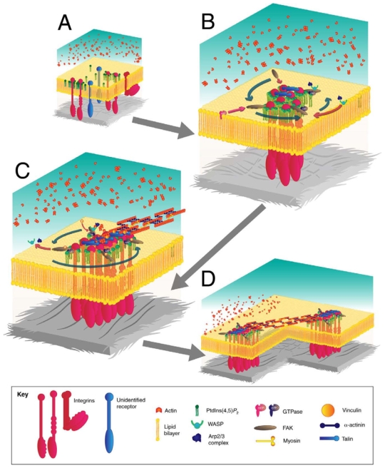
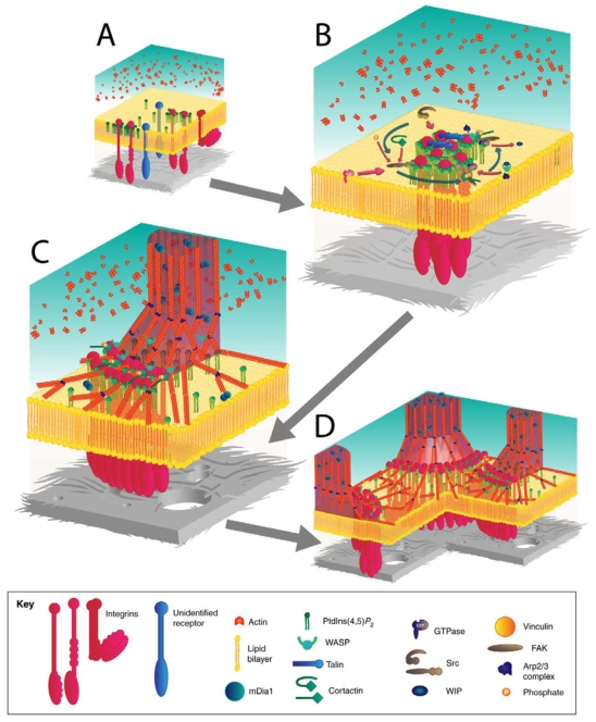
The invasiveness of cells is correlated with the presence of dynamic actin-rich membrane structures called invadopodia, which are membrane protrusions that are associated with localized polymerization of sub-membrane actin filaments. Similar to focal adhesions and podosomes, invadopodia are cell-matrix adhesion sites. Indeed, invadopodia share several features with podosomes, but whether they are distinct structures is still a matter of debate. Invadopodia are built upon an N-WASP-dependent branched actin network, and the Rho GTPase Cdc42 is involved in inducing invadopodial-membrane protrusion, which is mediated by actin filaments that are organized in bundles to form an actin core. Actin-core formation is thought to be an early step in invadopodium assembly, and the actin core is perpendicular to the extracellular matrix and the plasma membrane; this contrasts with the tangential orientation of actin stress fibers anchored to focal adhesions. In this Commentary, we attempt to summarize recent insights into the actin dynamics of invadopodia and podosomes, and the forces that are transmitted through these invasive structures. Although the mechanisms underlying force-dependent regulation of invadopodia and podosomes are largely unknown compared with those of focal adhesions, these structures do exhibit mechanosensitivity. Actin dynamics and associated forces might be key elements in discriminating between invadopodia, podosomes and focal adhesions. Targeting actin-regulatory molecules that specifically promote invadopodium formation is an attractive strategy against cancer-cell invasion.


Figures

Fig. 1
Fig. 1
Schematic view of signaling pathways that lead to actin organization at focal adhesions. (A) At the initial stage of adhesion formation, integrins or other unidentified receptors bind to components of the ECM (grey), leading to clustering of receptors into PtdIns(4,5)P2-enriched areas of plasma membrane. (B) In early spreading adhesions at the cell periphery, the Arp2/3 complex and WASP are targeted to adhesions by FAK. Blue arrows represent the spatiotemporal sequence of structure assembly. Pink arrows indicate protein recruitment. (C) Autophosphorylation of FAK at Tyr397 destabilizes the Arp2/3-WASP-FAK complex. Talin is recruited to adhesions, allowing integrin-ECM linkages to be functionally coupled to actomyosin; this enables actomyosin contractility to affect adhesion reinforcement and subsequent maturation. Actin filaments can be crosslinked by α-actinin. Myosin II incorporates into the α-actinin-crosslinked actin-filament bundles. (D) The collective dynamics of focal adhesions can be imaged by the actin-stress-fiber-mediated connection of focal adhesions. Connections can be observed between focal adhesions at the front of the cell with sliding trailing adhesions at the rear of the cell.
Fig. 2
Fig. 2
Schematic view of signaling pathways that lead to actin organization at invadopodia or podosomes. (A) At the initial stage of adhesion formation, integrins or other unidentified receptors bind to components of the ECM (grey), leading to clustering of receptors into PtdIns(4,5)P2-enriched areas of plasma membrane. (B) Recruitment of Src to adhesion sites leads to phosphorylation of several proteins such as cortactin, WASP, FAK and regulators of small GTPases. Continuous actin nucleation relies on the continuous and strong activation of the Arp2/3 complex at the membrane through the synergistic action of cortactin and WASP-family proteins. (C) DRF/mDia1 elongates actin filaments into columnar structures from the branched actin network that was previously induced by N-WASP, the Arp2/3 complex and cortactin. (D) Podosomes or invadopodia are mechanically connected through a network of radial actin filaments that lie parallel to the substratum.
Fig. 3
Fig. 3
Three-dimensional (3D) reconstruction of F-actin structure in a BHKRSV cell. (A) A 3D reconstruction was derived by combining images from confocal planes viewed from the side of the basal (adherent) face. F-actin staining was carried out after fixation in 4% paraformaldehyde with TRITCphalloidin. 3D reconstruction and rendering of the actin cytoskeleton was carried out through EDIT3D software, using grey-level images of each confocal z-stack (developed by Yves Usson and Franck Parazza, UMR CNRS 5525, Grenoble, France). Actin stress fibers are indicated by arrowheads, and the collective organization of podosomes and invadopodia by arrows. (B) A color scale was added, purely to indicate the relative position of the z-plane. The most basal plane was colored blue. Scale bars: 5 μm.
Fig. 4
Fig. 4
Actin dynamics in stress fibers and invadopodia rosettes in mouse embryonic fibroblasts transformed with Src. (A) Recovery of GFP-actin after photobleaching (green rectangles) is faster in invadopodial rosettes than in stress fibers. Images were extracted from a time series in which mouse embryonic fibroblasts expressing Src and GFP-actin were shown to form both invadopodial rosettes and stress fibers. Imaging and photobleaching conditions were exactly the same in both conditions. (B) Analysis of normalized fluorescence intensity shows that the net flux of actin, which is determined by the tangent at the origin of the recovery curve (black arrows), is faster in podosomes than in stress fibers. The plateau of the recovery curve does not reach the same level as before photobleaching, allowing the determination of the immobile fraction in each structure. From this analysis, it seems that stress fibers are composed mostly of poorly dynamic F-actin.
Fig. 5
Fig. 5
Visualization of actin structure and paxillin in mRFP-actin-transfected BHK-RSV cells. (A, B) Rigid (A) and flexible (B) substrates were coated with vitronectin. Staining for paxillin (green) was carried out after fixation in 4% paraformaldehyde with anti-paxillin antibodies. 3D reconstruction (right-most images) was carried out using EDIT3D software as in Fig. 3. (A) On the left is an image from a single confocal plane of a BHK-RSV cell adherent on glass (rigid) substrate. The right image shows a 3D reconstruction. Arrowheads indicate focal adhesions. (B) Left and middle panels show images from two confocal planes of a BHK-RSV cell adherent on hydrogel made of polyacrylamide (flexible) substrate. Confocal planes were from the top of the gel (left) and inside the gel (middle). Note that focal adhesions (arrowheads) are smaller in size on the flexible substrate (B) than on the rigid one (A). (B) The 3D reconstruction (right) shows that collective organizations of podosomes or invadopodia (arrow) seem to ‘push’ the gel, hauling the whole cell body.

References

    1. Abou-Kheir W, Isaac B, Yamaguchi H, Cox D. Membrane targeting of WAVE2 is not sufficient for WAVE2-dependent actin polymerization: a role for IRSp53 in mediating the interaction between Rac and WAVE2. J Cell Sci. 2008;121:379–390. - PMC - PubMed
    1. Abram CL, Seals DF, Pass I, Salinsky D, Maurer L, Roth TM, Courtneidge SA. The adaptor protein fish associates with members of the ADAMs family and localizes to podosomes of Src-transformed cells. J Biol Chem. 2003;278:16844–16851. - PubMed
    1. Alexander NR, Branch KM, Parekh A, Clark ES, Iwueke IC, Guelcher SA, Weaver AM. Extracellular matrix rigidity promotes invadopodia activity. Curr Biol. 2008;18:1295–1299. - PMC - PubMed
    1. Applewhite DA, Barzik M, Kojima S, Svitkina TM, Gertler FB, Borisy GG. Ena/VASP proteins have an anti-capping independent function in filopodia formation. Mol Biol Cell. 2007;18:2579–2591. - PMC - PubMed
    1. Arnold M, Cavalcanti-Adam EA, Glass R, Blummel J, Eck W, Kantlehner M, Kessler H, Spatz JP. Activation of integrin function by nanopatterned adhesive interfaces. Chemphyschem. 2004;5:383–388. - PubMed

Publication types