. 2009 Sep 1;122(Pt 17):3161-8.
doi: 10.1242/jcs.051193.

An N-terminal addressing sequence targets NLRX1 to the mitochondrial matrix

Affiliations

An N-terminal addressing sequence targets NLRX1 to the mitochondrial matrix

Damien Arnoult et al. J Cell Sci. .

Abstract

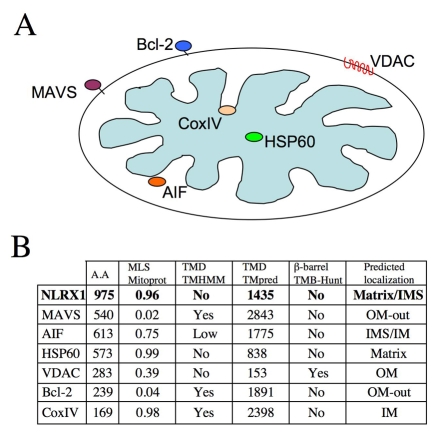
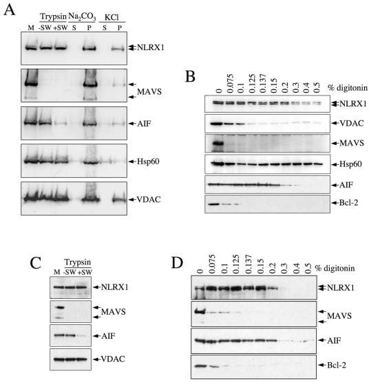
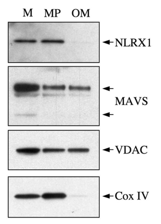
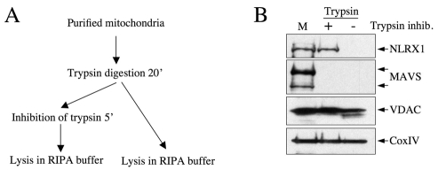
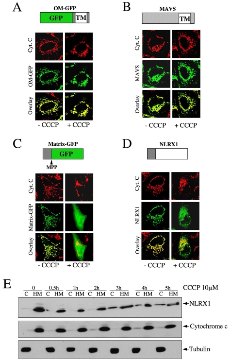
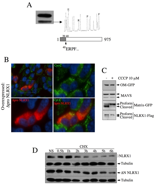
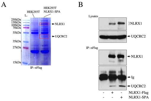
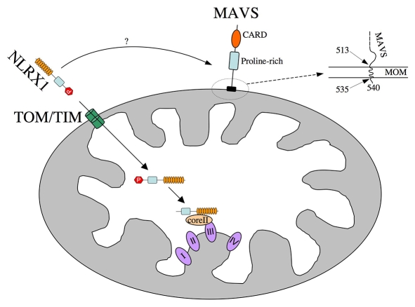
NLRX1 is the only member of the Nod-like receptor (NLR) family that is targeted to the mitochondria, and its overexpression induces the generation of reactive oxygen species (ROS), thus impacting on NFkappaB- and JNK-dependent signaling cascades. In addition, NLRX1 has been shown to interact with MAVS (also known as IPS-1, VISA and Cardif) at the mitochondrial outer membrane and to modulate antiviral responses. Here we report that NLRX1 has a functional leader sequence and fully translocates to the mitochondrial matrix via a mechanism requiring the mitochondrial inner-membrane potential, DeltaPsim. Importantly, we failed to detect NLRX1 at the mitochondrial outer membrane. We also show that the leader sequence of NLRX1 is removed, which generates a mature protein lacking the first 39 amino acids through a maturation process that is common for mitochondrial-matrix proteins. Finally, we identified UQCRC2, a matrix-facing protein of the respiratory chain complex III, as an NLRX1-interacting molecule, thus providing a molecular basis for the role of NLRX1 in ROS generation. These results provide the first identification of a protein belonging to the NLR family that is targeted to the mitochondrial matrix.


Figures

Fig. 1.
Fig. 1.
In silico analysis of the NLRX1 protein sequence. (A) Mitochondrial subcellular localization of MAVS, VDAC, Bcl-2, AIF, Cox IV and HSP60. (B) Algorithms were used to evaluate the presence of an N-terminal mitochondrial-addressing sequence (Mitoprot), transmembrane domains (TMHMM and TMpred) and β-barrels (TMB-Hunt). Numerical values computed by Mitoprot predict N-terminal mitochondrial-addressing sequences, ranging from 0 (lowest probability) to 1 (highest probability), and TMpred usually predicts a transmembrane domain for values >1750. Mitochondrial proteins of known subcellular localization (MAVS, AIF, HSP60, VDAC, Bcl-2, Cox IV) were also analyzed, to evaluate the strength of the algorithms. The resulting predicted subcellular localization was deduced from these analyses.
Fig. 2.
Fig. 2.
NLRX1 is a mitochondrial-matrix protein. (A) Purified mitochondria (M) from HEK293T cells overexpressing NLRX1-FLAG were treated with trypsin, in regular buffer (–SW) or in swelling conditions (+SW), or treated with Na2CO3 or KCl prior to centrifugation. S, supernatant; P, pellet. The resulting protein samples were analyzed by western blot with antibodies against FLAG, or against several mitochondrial markers, as indicated. (B) Purified mitochondria from HEK293T cells overexpressing NLRX1-FLAG were treated with increasing concentrations of digitonin, as indicated, and the pellet after centrifugation was analyzed by western blot for NLRX1, using an anti-FLAG antibody. Other mitochondrial markers were also analyzed, as indicated. (C) Purified mitochondria (M) from HEK293T cells were treated with trypsin in regular buffer (–SW) or in swelling conditions (+SW) and the presence of endogenous NLRX1 was determined by western blot using an anti-NLRX1 antibody, together with other mitochondrial markers, as indicated. (D) As in B but without transfection, and the indicated endogenous proteins were analyzed.
Fig. 3.
Fig. 3.
NLRX1 is undetectable at the mitochondrial outer membrane. Purified mitochondria (M) were fractioned by ultracentrifugation into mitoplasts (MP), which are enriched in inner-membrane proteins but still contain outer-membrane proteins, and pure outer membrane (OM). The presence of the endogenous NLRX1, as well as known mitochondrial markers, was analyzed by western blotting.
Fig. 4.
Fig. 4.
NLRX1 is not an integral MIM protein. (A) Schematic representation of the procedure applied. (B) Purified mitochondria (M) were treated with trypsin for 20 minutes and then lysed in RIPA buffer in the presence (+) or absence (–) of a trypsin inhibitor. The presence of endogenous NLRX1, as well as of known mitochondrial markers, was analyzed by western blotting.
Fig. 5.
Fig. 5.
The import of NLRX1 into mitochondria requires the ΔΨm. (A-D) HeLa cells were grown on coverslips, transfected overnight with OM-GFP (A), FLAG-MAVS (B), Matrix-GFP (C) or NLRX1-FLAG (D) and incubated or not with 10 μM CCCP for 8 hours before fixation and analysis by immunofluorescence. Mitochondria were visualized using an anti-cytochrome-c antibody. MAVS and NLRX1 were visualized using an anti-FLAG antibody. (E) HeLa cells transfected with NLRX1-FLAG were incubated for various times with 10 μM CCCP, and cytosol (C) and heavy membrane (HM) that contained mitochondria were analyzed by western blotting, using antibodies against FLAG (for NLRX1), cytochrome c and tubulin.
Fig. 6.
Fig. 6.
The N-terminal leader sequence of NLRX1 is cleaved following transport. (A) The identity of the faster migrating band of NLRX1 was determined, after membrane excision, by Edman degradation, revealing maturation between amino acids 39 and 40 of the NLRX1 prosequence. (B) A truncated form of NLRX1, lacking the (1-39) prosequence, is unable to localize to mitochondria, as determined by immunofluorescence. Mitochondria and Δpro-NLRX1–FLAG were visualized using anti-cytochrome-c and anti-FLAG antibodies, respectively. (C) HeLa cells transfected and stimulated with 10 μM CCCP, as in Fig. 2, were analyzed by western blot with the indicated antibodies. (D) HeLa cells were transfected with either NLRX1-FLAG or ΔN-NLRX1–FLAG, treated with 100 μM cycloheximide (CHX) for the indicated times, and cell lysates analyzed by western blot using an anti-FLAG antibody. Membranes were stripped and reblotted with an anti-tubulin antibody for normalization.
Fig. 7.
Fig. 7.
A free N-terminal NLRX1 addressing sequence is necessary and sufficient for mitochondrial targeting. (A,C) HeLa cells were grown on coverslips, transfected with NLRX1(1-156)-GFP (A) or HA-NLRX1 (C), fixed and analyzed by immunofluorescence using anti-cytochrome-c (A,C), anti-NLRX1 and anti-HA (C), as indicated. (B,D) HEK293T cells were transfected with NLRX1(1-156)-GFP (B) or HA-NLRX1 (D) and analyzed by western blotting using anti-GFP (B), anti-NLRX1 and anti-HA (D), as indicated.
Fig. 8.
Fig. 8.
Pulldown assays identify UQCRC2 as a binding partner of NLRX1. (A) Lysates from HEK293T and HEK293T NLRX1-SPA cell lines were immunoprecipitated using a monoclonal anti-FLAG antibody and bound proteins were resolved by SDS-PAGE. Bands found only in immunoprecipitates from HEK293T NLRX1-SPA cells were identified by mass spectrometry. L, ladder. (B) HEK293T cells were transfected with NLRX1-FLAG or NLRX1-SPA. Cell lysates were analyzed using anti-FLAG and anti-UQCRC2 antibodies. Lysates were immunoprecipitated using anti-UQCRC2 antibody and analyzed by western blot using anti-FLAG and anti-UQCRC2 antibodies. Ig, immunoglobulins.
Fig. 9.
Fig. 9.
Schematic model for the internalization of NLRX1 into the mitochondrial matrix. Schematic representation of the proposed steps for the import of NLRX1 to the matrix face of the inner membrane. MAVS is also shown in this representation, and the precise insertion of the MAVS C-terminal sequence into the MOM is illustrated, and is derived from the study by Seth et al. (Seth et al., 2005). The four complexes (I-IV) of the mitochondrial respiratory chain are shown, as well as the core II protein (UQCRC2) of the complex III (bc1 complex). P, prosequence (aa 1-39); CARD, caspase-activation and -recruitment domain.

References

    1. Akira, S., Uematsu, S. and Takeuchi, O. (2006). Pathogen recognition and innate immunity. Cell 124, 783-801. - PubMed
    1. Arnoult, D. (2008). Apoptosis-associated mitochondrial outer membrane permeabilization assays. Methods 44, 229-234. - PubMed
    1. Bell, E. L., Klimova, T. A., Eisenbart, J., Moraes, C. T., Murphy, M. P., Budinger, G. R. and Chandel, N. S. (2007). The Qo site of the mitochondrial complex III is required for the transduction of hypoxic signaling via reactive oxygen species production. J. Cell Biol. 177, 1029-1036. - PMC - PubMed
    1. Benko, S., Philpott, D. J. and Girardin, S. E. (2008). The microbial and danger signals that activate Nod-like receptors. Cytokine 43, 368-373. - PubMed
    1. Fritz, J. H., Ferrero, R. L., Philpott, D. J. and Girardin, S. E. (2006). Nod-like proteins in immunity, inflammation and disease. Nat. Immunol. 7, 1250-1257. - PubMed

Publication types

MeSH terms