Comparative Study
. 2009 Aug 19;29(33):10299-308.
doi: 10.1523/JNEUROSCI.2500-09.2009.

Sonic hedgehog pathway activation is induced by acute brain injury and regulated by injury-related inflammation

Affiliations
Comparative Study

Sonic hedgehog pathway activation is induced by acute brain injury and regulated by injury-related inflammation

Nduka M Amankulor et al. J Neurosci. .

Abstract

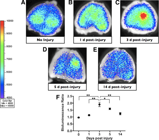
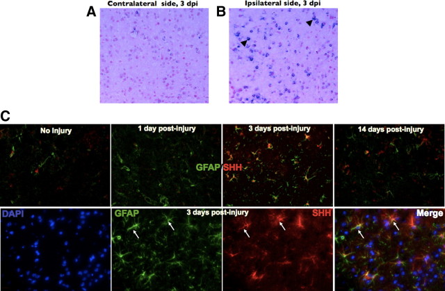
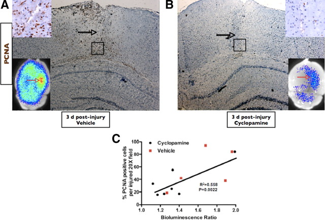
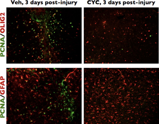
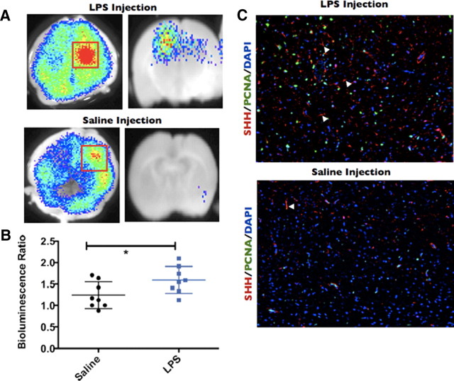
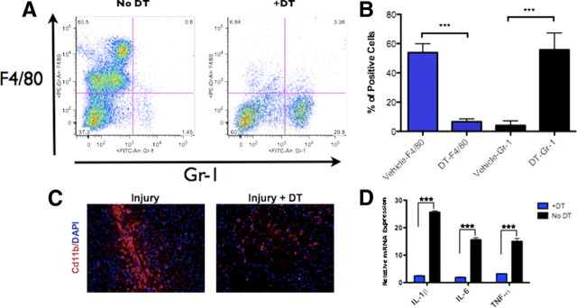
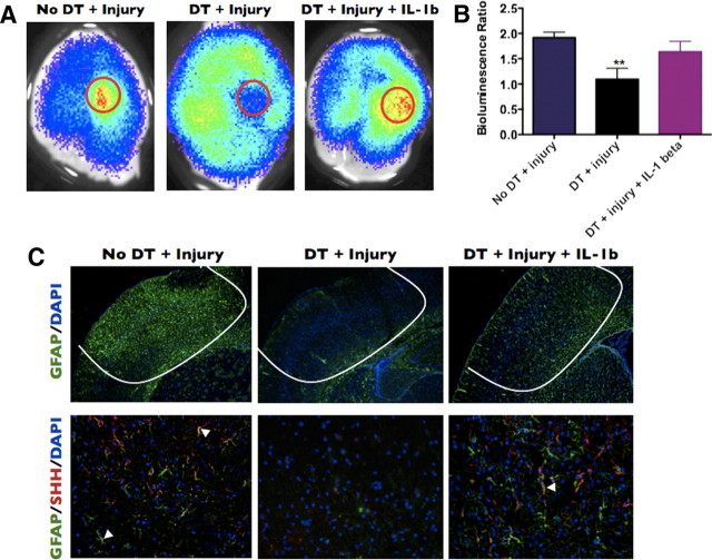
The adult mammalian brain responds to injury by activating a program of cell proliferation during which many oligodendrocyte precursors, microglia, and some astrocytes proliferate. Another common response to brain injury is the induction of reactive gliosis, a process whereby dormant astrocytes undergo morphological changes and alter their transcriptional profiles. Although brain injury-induced reactive gliosis is concurrent with the proliferation of surrounding cells, a functional relationship between reactive gliosis and this cell proliferation has not been clearly demonstrated. Here, we show that the mitogen sonic hedgehog (SHH) is produced in reactive astrocytes after injury to the cerebral cortex and participates in regulating the proliferation of Olig2-expressing (Olig2(+)) cells after brain injury. Using a cortical freeze injury to induce reactive gliosis in a Gli-luciferase reporter mouse, we show that the SHH pathway is maximally active 3 d after brain injury and returns to baseline levels by 14 d. SHH expression parallels Gli activation and localizes to glial fibrillary acidic protein-expressing reactive astrocytes. Inhibition of the SHH pathway with cyclopamine blocks the Gli response and significantly reduces both the proliferating and overall number of Olig2(+) cells in the injured cortex. To provide mechanistic insight into SHH pathway activation in astrocytes, we show that proinflammatory stimuli activate SHH-expressing reactive astrocytes, whereas inhibition of inflammation-induced reactive gliosis by macrophage depletion abolishes SHH activation after brain injury and dampens cell proliferation after injury. Our data describes a unique reactive astrocyte-based, SHH-expressing niche formed in response to injury and inflammation that regulates the proliferation of Olig2(+) cells.


Figures

Figure 1.
Figure 1.
The Gli pathway is activated after a cortical freeze injury in the mouse brain. A–E, Gli bioluminescence imaging of an uninjured brain and brains imaged 1, 3, 5, and 14 d after injury, respectively. A focal increase in BLI intensity was noted at 3 and 5 d after injury. F, Quantification and statistical analysis of the BLI intensity over the injury area compared with the identical area in the uninjured cortex of the same mouse.
Figure 2.
Figure 2.
SHH is expressed in the mouse brain after brain injury. A, B, In situ hybridization for SHH in the uninjured contralateral hemisphere and injured brain, respectively. Black arrowheads indicate positively stained cells (blue) in the injured right cortex. C, Top, from left to right, SHH and GFAP double immunofluorescence from uninjured brain and brains processed after 1, 3, and 14 d after injury, respectively. C, Bottom, from left to right, High-magnification immunofluorescence staining of a mouse brain at 3 d after injuring showing 4′,6′-diamidino-2-phenylindole (DAPI), GFAP, SHH, and a merged image, respectively. White arrows indicate astrocyte costaining for SHH and GFAP. Images in A, B, and C (top) were taken at 20× magnification. Images in C (bottom) were taken at 63× magnification. dpi, Days post-injury.
Figure 3.
Figure 3.
Cyclopamine disrupts Gli activation after brain injury. A–C, Gli–Luc bioluminescence was obtained in vehicle-treated and uninjured mice, vehicle-treated and injured mice (3 d after injury), and cyclopamine-treated and injured mice (3 d after injury). D, Quantification of Gli–Luc bioluminescence for A–C. Letters in the bar graph correspond to the BLI images. p < 0.01 for vehicle-treated and injured mice compared with either uninjured or cyclopamine-treated mice.
Figure 4.
Figure 4.
Cyclopamine reduces the proliferative capacity of injured brain. A, B, PCNA and BLI of cyclopamine-treated and vehicle-treated brains at 3 d after injury, respectively. Open black arrows point to the needle tracks for each injury. Black boxes identify the area magnified in the top inset of each image. The bottom insets are BLI images of the brains that correspond to the immunohistochemistry micrograph. Open red arrows point to the area of injury for the BLI images. C, Simple linear regression showing a positive correlation (r2) between Gli BLI intensity and PCNA expression in injured brains and the corresponding p value. Both vehicle-treated (red squares; n = 5) and cyclopamine-treated (black circles; n = 7) mice were included in the regression analysis. A and B were taken under 5× magnification; inset PCNA images were taken under 40× magnification.
Figure 5.
Figure 5.
Cyclopamine preferentially reduces the number of proliferating Olig2 cells after brain injury. Brains from cyclopamine-treated (CYC) or vehicle-treated (Veh) mice injured 3 d before the animals were killed were used for immunofluorescence with an antibody to PCNA and antibodies to either Olig2 or GFAP. All images taken at 10× magnification.
Figure 6.
Figure 6.
Intracerebral injection of LPS is capable of inducing Gli activation and SHH expression. Gli–Luc BLI of LPS-injected (A, top) or saline-injected (A, bottom) mouse brains processed 3 d after injection. Red squares indicate the area of Gli activation. Left column is whole-brain BLI images, whereas right column is coronal slices made at the point of injury. Note that saline-injected brains also display increased Gli activation in the region of needle penetration. B, Column scatter graph showing the bioluminescence ratio of saline-injected (black circles) or LPS-injected (blue squares) brains. C, SHH, PCNA, and DAPI immunofluorescence staining for LPS-injected (top) or saline-injected (bottom) brains. Filled white arrowheads indicate SHH-positive cells. Images taken at 10× magnification. *p < 0.05.
Figure 7.
Figure 7.
Treating CD11b–DTR mice with DT reduces the total number of peritoneal and brain macrophages and induces a marked reduction in cytokine production after brain injury. A, Representative flow cytometry plot of cells obtained from peritoneal lavage of injured CD11b–DTR mice treated with DT or vehicle alone after staining for F4/80 (macrophage marker) and Gr-1 (neutrophil marker). There is a reduction in F4/80-positive cells (macrophages), as quantified in B (***p < 0.001). C, Immunofluorescence staining for CD11b and DAPI in injured brains from CD11b–DTR mice (3 d after injury) treated with or without DT. D, Quantitative RT-PCR analysis of injured brains (3 d after injury) from mice treated with DT or vehicle alone (***p < 0.001). Images in C taken at 20× magnification.
Figure 8.
Figure 8.
Macrophage depletion reduces Gli activation, astroglial activation, and SHH expression in brain-injured mice. A, CD11b-DTR mice injected with vehicle, DT, or with DT plus an intracerebral injection of mouse IL-1β were killed 3 d after cortical freeze injury, and BLI images were obtained. B, Quantification of the bioluminescence ratio in A. C, Immunofluorescence of a brain from the same groups as in A stained for either GFAP and DAPI (top row) or GFAP, SHH, and DAPI (bottom row). C, Top row, 5× magnification; bottom row, 20× magnification. **p < 0.01.

References

    1. Ahn S, Joyner AL. In vivo analysis of quiescent adult neural stem cells responding to Sonic hedgehog. Nature. 2005;437:894–897. - PubMed
    1. Alderson P, Roberts I. Corticosteroids for acute traumatic brain injury. Cochrane Database Syst Rev. 2005;2005:CD000196. - PMC - PubMed
    1. Androutsellis-Theotokis A, Leker RR, Soldner F, Hoeppner DJ, Ravin R, Poser SW, Rueger MA, Bae SK, Kittappa R, McKay RD. Notch signalling regulates stem cell numbers in vitro and in vivo. Nature. 2006;442:823–826. - PubMed
    1. Becher OJ, Hambardzumyan D, Fomchenko EI, Momota H, Mainwaring L, Bleau AM, Katz AM, Edgar M, Kenney AM, Cordon-Cardo C, Blasberg RG, Holland EC. Gli activity correlates with tumor grade in platelet-derived growth factor-induced gliomas. Cancer Res. 2008;68:2241–2249. - PubMed
    1. Buffo A, Vosko MR, Ertürk D, Hamann GF, Jucker M, Rowitch D, Götz M. Expression pattern of the transcription factor Olig2 in response to brain injuries: implications for neuronal repair. Proc Natl Acad Sci U S A. 2005;102:18183–18188. - PMC - PubMed

Publication types

MeSH terms

Substances

LinkOut - more resources