. 2009 Sep 15;183(6):3720-30.
doi: 10.4049/jimmunol.0900970. Epub 2009 Aug 19.

Tumor-derived microvesicles promote regulatory T cell expansion and induce apoptosis in tumor-reactive activated CD8+ T lymphocytes

Affiliations

Tumor-derived microvesicles promote regulatory T cell expansion and induce apoptosis in tumor-reactive activated CD8+ T lymphocytes

Eva U Wieckowski et al. J Immunol. .

Abstract

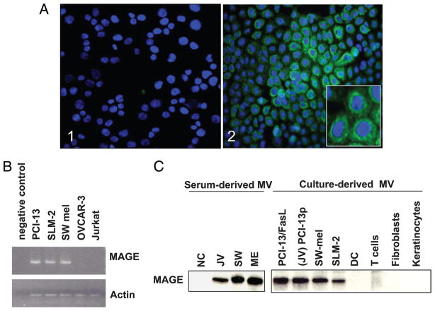
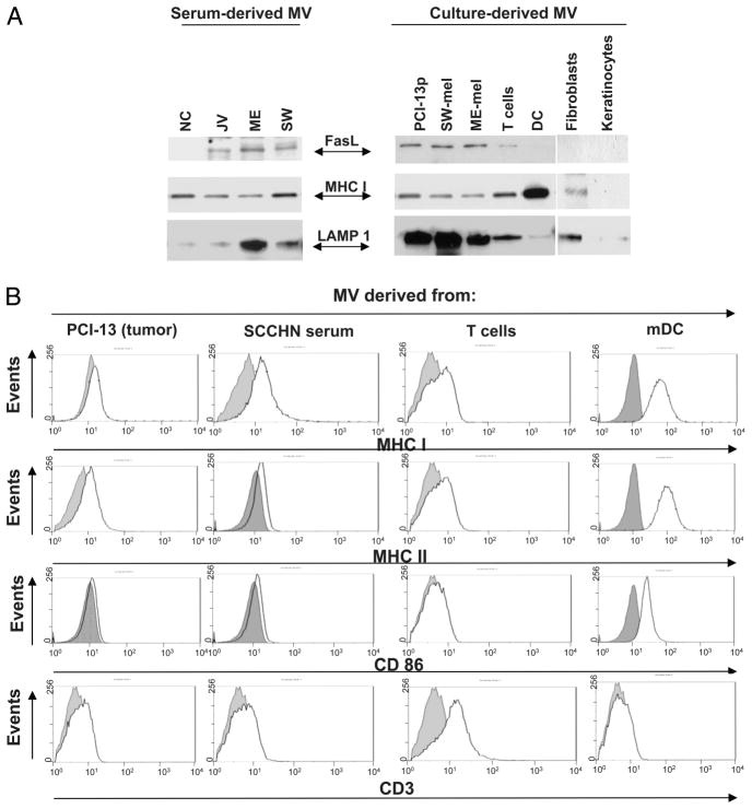
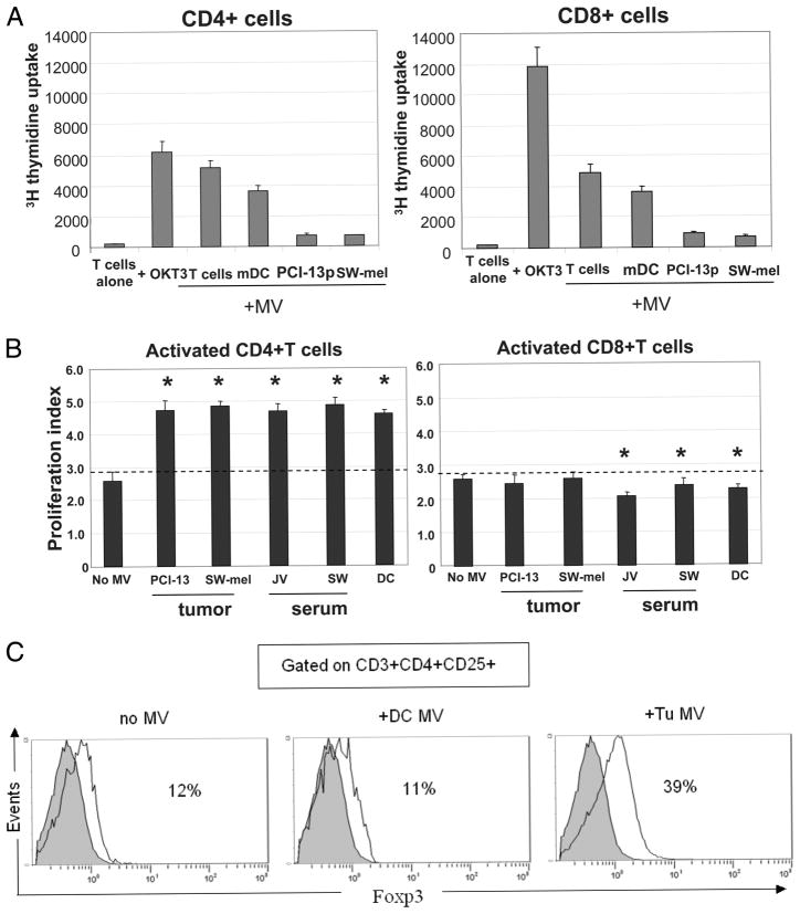
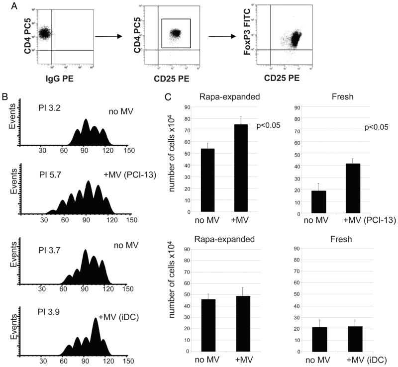
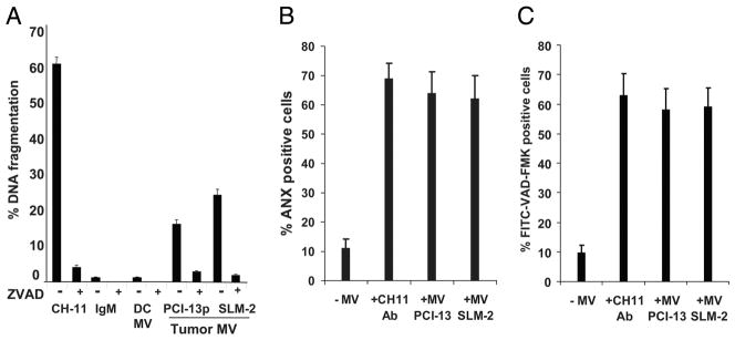
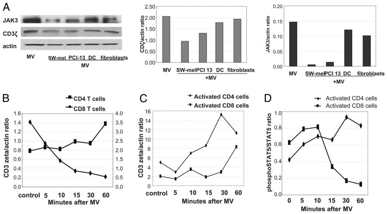
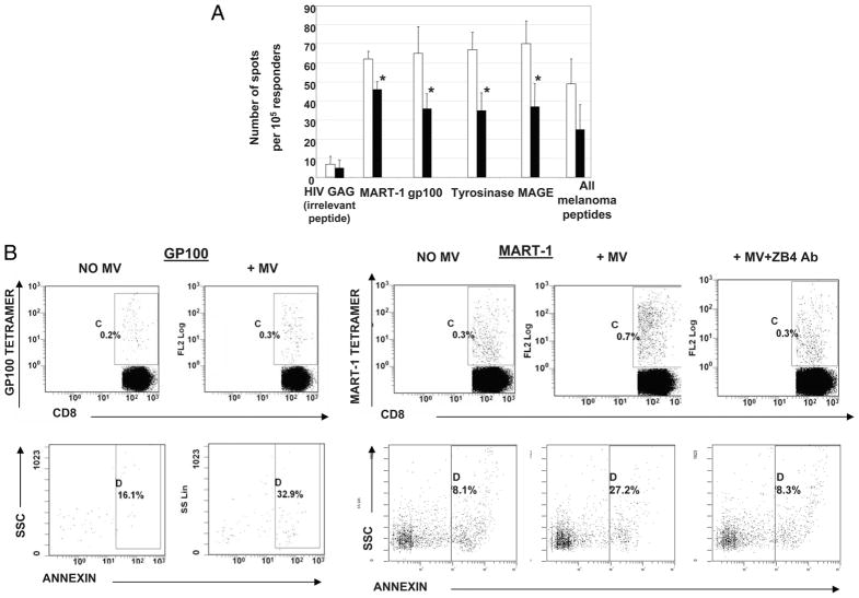
Sera of patients with cancer contain membraneous microvesicles (MV) able to induce apoptosis of activated T cells by activating the Fas/Fas ligand pathway. However, the cellular origin of MV found in cancer patients' sera varies as do their molecular and cellular profiles. To distinguish tumor-derived MV in cancer patients' sera, we used MAGE 3/6(+) present in tumors and MV. Molecular profiles of MAGE 3/6(+) MV were compared in Western blots or by flow cytometry with those of MV secreted by dendritic cells or activated T cells. These profiles were found to be distinct for each cell type. Only tumor-derived MV were MAGE 3/6(+) and were variably enriched in 42-kDa Fas ligand and MHC class I but not class II molecules. Effects of MV on signaling via the TCR and IL-2R and proliferation or apoptosis of activated primary T cells and T cell subsets were also assessed. Functions of activated CD8(+) and CD4(+) T lymphocytes were differentially modulated by tumor-derived MV. These MV inhibited signaling and proliferation of activated CD8(+) but not CD4(+) T cells and induced apoptosis of CD8(+) T cells, including tumor-reactive, tetramer(+)CD8(+) T cells as detected by flow cytometry for caspase activation and annexin V binding or by DNA fragmentation. Tumor-derived but not dendritic cell-derived MV induced the in vitro expansion of CD4(+)CD25(+)FOXP3(+) T regulatory cells and enhanced their suppressor activity. The data suggest that tumor-derived MV induce immune suppression by promoting T regulatory cell expansion and the demise of antitumor CD8(+) effector T cells, thus contributing to tumor escape.


Conflict of interest statement

Disclosures

The authors have no financial conflict of interest.

Figures

FIGURE 1
FIGURE 1
MAGE 3/6 is expressed by tumor cells and is present on tumor-derived MV. A, PCI 13 tumor cells immunostained for MAGE 3/6: 1) isotype control; and 2) a confocal microscopy image showing intracytoplasmic staining (green) for MAGE 3/6; cell nuclei are blue (4′,6-diamidino-2-phenylindole (DAPI+)); magnification, ×200. Inset, magnification, ×600. B, Expression of MAGE 3/6 by tumor cells as tested by RT-PCR. A representative of three experiments performed. C, Western blot analysis of MAGE 3/6 expression in serum-derived or cell culture-derived MV. A representative of five Western blot experiments performed is shown.
FIGURE 2
FIGURE 2
Western blot and flow cytometry analyses of MV derived from tumor cells, sera of patients with cancer, activated T cells, mDC, and cultured tumor or normal cells. A, Western blots of serum derived or culture-derived MV. Each lane was loaded with 25 μg of protein. A representative of 10 Western blot experiments performed is shown. B, Flow analysis of MV bound to latex beads. Aliquots of bead-bound MV were incubated with the PE-labeled Abs as indicated. Dotted lines indicate controls (i.e., bead-bound MV incubated with isotype control). A representative experiment of six performed is shown.
FIGURE 3
FIGURE 3
Effects of MV on proliferation of CD4+ or CD8+ T lymphocyte subsets. A, Resting CD4+ and CD8+ T cells were purified and coin-cubated with MV of various origins. Proliferation was measured by [3H]thy-midine incorporation. Responder cells (0.5 × 10⁶) were cultured in the presence of MV (200 μg of protein). MV were isolated from activated T cells, mDC, SW-mel, or PCI-13 cells. As a positive control, OKT-3 Ab was used. B, Effects of MV on proliferation of activated CD4+ or CD8+ T cells. MV of different origins were coincubated with CFSE-labeled, OKT-3 Ab-activated T cell subsets, and proliferation was assayed by flow cytometry. The proliferation index was calculated using Modfit software. The data are mean values ± SD. The asterisks indicate p < 0.001 for CD4+ T cells and p < 0.01 for CD8+ T cells. C, Four-color flow cytometry analysis of activated CD4+ T cell populations cultured in the presence or absence of tumor-derived (PCI-13) or DC-derived MV. On day 7 of culture, cells were tested for CD25 and FOXP3 expression. The percentages of FOXP3+ cells within the CD3+CD4+ CD25+ subset of cells are indicated in each panel. The data shown in A–C are from individual representative experiments of five performed with T cells obtained from different normal donors.
FIGURE 4
FIGURE 4
Tumor-derived MV expand Treg. Isolated CD4+CD25+ T cells were cultured in the presence of rapamycin as described in Materials and Methods. A, Phenotypic characteristics of rapamycin-expanded Treg tested in day 20 of culture. All expanding cells were CD4+CD25highFOXP3+. B, Proliferation of CFSE-labeled, ex vivo-generated Treg ± MV in 7-day cultures. MV were either tumor-derived (PCI-13) or were isolated from supernatants of human iDC. Proliferation index (PI) for each culture was obtained using Modfit software. C, Cell numbers (means ± SD of 4 wells) in cultures of rapamycin (Rapa)-expanded (left) or freshly isolated (right) Treg incubated ± MV derived from PCI-13 or iDC supernatants. The data shown in C are from three independent experiments.
FIGURE 5
FIGURE 5
Tumor-derived MV induce apoptosis and signaling defects in Jurkat cells. A, Apoptosis of CD8+ Jurkat cells after coincubation with MV measured in JAM assay as DNA fragmentation. Apoptosis was blocked by preincubation of T cells with pan-caspase inhibitor Z-VAD-FMK. Data are means ± SD of quadruplicate wells obtained in 10 experiments performed is shown. B, A representative experiment of caspase activation in CD8+ Jurkat cells coincubated with tumor-derived MV or CH-11A or Ab isotype control for 24 h. C, Annexin V (ANX) binding to CD8+ Jurkat cells coincubated with tumor-derived MV as in B. The data in B and C are mean values ± SD from one representative experiment of five performed.
FIGURE 6
FIGURE 6
MV induce down-regulation of CD3ζ and JAK3 expression in primary-activated CD8+ T cells. A, Western blot analysis of CD3ζ and JAK3 expression in activated T cells after coincubation with MV. The CD3ζ/actin and JAK3/actin ratios for the Western blot analysis are shown on the right. The data are representative of five experiments performed with primary T cells of different donors. B, CD3ζ expression in nonactivated (resting) T cells ± MV. C, CD3ζ expression in the T cell subsets activated with microbeads coated with OKT3 and anti-CD28 mAb for 48 h before the addition of MV. The data in B and C are ratios of CD3ζ/actin derived from Western blot analyses performed at different times after tumor-derived MV were added to activated T cells. D, Phosphorylated STAT5/STAT5 ratios in T cell subsets activated as indicated above and incubated ± MV. BD, Representative experiments of three performed from each condition is shown.
FIGURE 7
FIGURE 7
Effects of MV on tumor peptide-reactive T cells. A, IFN-γ ELISPOT analysis of an HLA-A2+ melanoma patient’s T cells generated by IVS with melanoma peptides presented by autologous mDC. These T cells were then coincubated with tumor-derived MV as described in Materials and Methods. The data are mean spot numbers ± SD from quadruplicate wells in a representative ELISPOT assay out of five performed. The numbers of IFN-γ spots produced by T cells not treated with MV (□) and (■) the numbers of spots produced by MV-treated T cells. MV-treated or MV-untreated T cells were stimulated with T2 cells loaded with the indicated peptides in ELISPOT assays. Asterisks indicate p < 0.01. B, Annexin V binding to melanoma-specific (MART-1 tetramer+ or gp100 tetramer+) T cells coincubated with tumor-derived MV or control MART-1 tetramer+ or gp100 tetramer+CD8+ T cells incubated in the absence of MV. The last right panel shows inhibition annexin V binding in the presence of ZB4 mAb. A representative experiment of three performed is shown.

References

    1. Denzer K, Kleijmeer MJ, Heijnen HF, Stoorvogel W, Gellze HJ. Exosome: from internal vesicle of the multivesicular body to intercellular signaling device. J Cell Sci. 2000;113:3365–3374. - PubMed
    1. Johnstone RM. Exosomes biological significance: a concise review. Blood Cells Mol Dis. 2006;36:315–321. - PubMed
    1. van Niel G, Porto-Carreiro I, Simors S, Raposo G. Exosomes: a common pathway for a specialized function. J Biochem. 2006;140:13–21. - PubMed
    1. Ratajczak J, Wysoczynski M, Hayek F, Janowska-Wieczorek A, Ratajczak MZ. Membrane-derived microvesicles: important and underappreciated mediators of cell-to-cell communication. Leukemia. 2006;20:1487–1495. - PubMed
    1. Martinez-Lorenzo MJ, Anel A, Gamen S, Monle NI, Lasierra P, Larrad L, Pineiro A, Alava MA, Naval J. Activated human T cells release bioactive Fas ligand and APO2 lignad in microvesicles. J Immunol. 1999;163:1274–1281. - PubMed

Publication types

MeSH terms