Review
. 2009 Sep;10(9):943-8.
doi: 10.1038/ni.1781. Epub 2009 Aug 19.

Foamy macrophages and the progression of the human tuberculosis granuloma

Affiliations
Review

Foamy macrophages and the progression of the human tuberculosis granuloma

David G Russell et al. Nat Immunol. 2009 Sep.

Abstract

The progression of tuberculosis from a latent, subclinical infection to active disease that culminates in the transmission of infectious bacilli is determined locally at the level of the granuloma. This progression takes place even in the face of a robust immune response that, although it contains infection, is unable to eliminate the bacterium. The factors or environmental conditions that influence this progression remain to be determined. Recent advances have indicated that pathogen-induced dysregulation of host lipid synthesis and sequestration serves a critical role in this transition. The foamy macrophage seems to be a key participant in both sustaining persistent bacteria and contributing to the tissue pathology that leads to cavitation and the release of infectious bacilli.


Figures

Box 1
Box 1. Histological Progression of the Human Tuberculosis Granuloma
The early events following infection with Mycobacterium tuberculosis appear relatively consistent. Infection is initiated following inhalation of viable bacilli present in exhaled droplets or nuclei that are discharged into the atmosphere by an individual with an active infection. These droplets can persist in the atmosphere for several hours and, because the infectious dose is in the range of 1-10 bacilli, this makes transmission an extremely efficient process. Once in the lung the bacilli are phagocytosed by alveolar macrophages. Internalization of the bacilli triggers a proinflammatory response that induces the macrophage to invade the subtending epithelium. This response also leads to recruitment of mononuclear cells from neighboring blood vessels. These monocytes form the cellular matrix of the early granuloma, which is the primary characteristic of this disease. In its early stage, the granuloma has a core of infected macrophages enclosed by foamy macrophages and other mononuclear phagocytes, surrounded by lymphocytes. This tissue response contains the infection and spells the end of the period of rapid replication for Mtb. As the granuloma matures it develops an extensive fibrous capsule that encases the macrophage core and excludes the majority of lymphocytes from the center of the structure. Concomitant with this transition is a marked reduction in the number of blood vessels penetrating the granuloma. At this stage there is a noticeable increase in the number of foamy macrophages within the fibrous capsule. We hypothesize that these cells are responsible for the accumulation of caseous debris in the center of the granuloma, which portends progression to active disease. In an immunocompetent person this progression is localized to individual granulomas and the same tissue site will harbor other granulomas that appear under perfect immune containment. Nonetheless, in a progressive infection the caseous, necrotic center of the granuloma liquefies and cavitates spilling thousands of infectious Mtb into the airways. This damage to the lungs triggers development of a productive cough facilitating generation of the infectious aerosol and completion of the bacterium's life cycle.
Figure 1
Figure 1
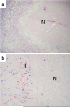
Within TB patients’ lesions, foamy macrophages are mainly located within the interface region surrounding central necrosis. Adapted from Peyron et al. (a). Typical necrotic lesion from a thin section made through a lymph node biopsy from a TB patient, stained with Haematoxylin and Eosin. As classically observed in human TB lesions, the lesion is well-circumscribed and differentiated with the central necrotic core (N) surrounded by a thin area called Interface (I) separating the necrosis from the histiocytes area. At the periphery, the lymphocyte area delineates the boundary of the structure. (b). Oil red-O staining of such TB lesions demonstrated the presence of large amounts of foamy macrophages within the interface region (I) separating necrosis (N) from the histiocytes, as shown in this enlarged views.
Figure 2
Figure 2
Vesicles containing Mycobacterium-derived cell wall lipids are released from infected macrophages. (a) Live macrophages infected for 24 h with Texas Red hydrazide-labeled Mycobacterium bovis BCG were analyzed by fluorescence microscopy, with striking release of Texas Red label from the bacterial phagosome. (b) These macrophages were incubated with dextran-fluorescein for 1 h demonstrating that the released lipids permeated the host macrophage and colocalized with dextran-fluorescein, revealing the penetrance of the endocytic system by the bacterial lipids. (c) Immunoelectron micrograph of an infected macrophage probed with Ab's against Mtb cell wall glycolipids PIM (anti-mouse IgM-12 nm gold) and LAM (anti rabbit IgG–18 nm gold). Labeling of bacterial lipids can be seen in multivesicular structures (arrowed, b=bacteria). (d). Platinum replicas revealing the surface of BCG-infected macrophages. BCG cell wall lipid-containing vesicles are exocytosed from macrophages infected with biotin hydrazide-labeled BCG. The presence of bacterial components were detected with Streptavidin-gold (15 nm, arrowed). (e) Shows a high magnification micrograph demonstrating the release of these vesicles at the cell surface. Notice that the cell membrane is free from label, which is restricted to the exocytosed vesicles. Panels (a), (b), and (d) are reproduced, with permission, from reference .
Figure 3
Figure 3
Within foamy macrophages, tubercle bacilli-containing phagosomes display privileged contact with cellular lipid bodies. (a). Tight apposition between Mtb-containing phagosomes and host lipid bodies (LB) can be observed (arrow). (b) Ultimately, at later stages, bacilli-containing phagosomes are observed within lipid bodies (LB), thus confirming that they have been engulfed. (c). Engulfed bacilli display intra-cytoplasmic lipid inclusions typical of dormant bacteria (ILI), which suggests strongly that they have accumulated host lipids.
Figure 4
Figure 4
A model for caseum accumulation and granuloma progression (a) Intracellular Mtb synthesize and release cell wall components inside their host cell. These lipids accumulate in the internal vesicles in multi-vesicular bodies, which are exocytosed from the cell in vesicular form. (b). Because of the release of these vesicles, both infected and uninfected macrophages are exposed to cell wall mycolates and induced to form foam cells. (c) These cells die via an inflammatory, necrotic process and release their lipid droplets into the extracellular milieu within the granuloma. (d). As a result of the fibrotic capsule the human granuloma is an enclosed, isolated structure. The enclosed nature of the human granuloma leads to accumulation of necrotic debris as caseum. We propose that this process is an integral part of the pathology that leads to active disease and transmission.

References

    1. Parrish NM, Dick JD, Bishai WR. Mechanisms of latency in Mycobacterium tuberculosis. Trends Microbiol. 1998;6:107–112. - PubMed
    1. Rohde K, Yates RM, Purdy GE, Russell DG. Mycobacterium tuberculosis and the environment within the phagosome. Immunol Rev. 2007;219:37–54. - PubMed
    1. Sturgill-Koszycki S, et al. Lack of acidification in Mycobacterium phagosomes produced by exclusion of the vesicular proton-ATPase. Science. 1994;263:678–81. - PubMed
    1. Gohn A, editor. Der primäre Lungenherd bei der Tuberkulose der Kinder (The Primary Lung Lesion in Infant TB) Berlin: 1912.
    1. Saunders BM, Cooper AM. Restraining mycobacteria Role of granulomas in mycobacterial infections. Immunol Cell Biol. 2000;78:334–341. - PubMed

Publication types