. 2009 Sep 3;461(7260):109-13.
doi: 10.1038/nature08268. Epub 2009 Aug 19.

Antioxidant and oncogene rescue of metabolic defects caused by loss of matrix attachment

Affiliations

Antioxidant and oncogene rescue of metabolic defects caused by loss of matrix attachment

Zachary T Schafer et al. Nature. .

Abstract

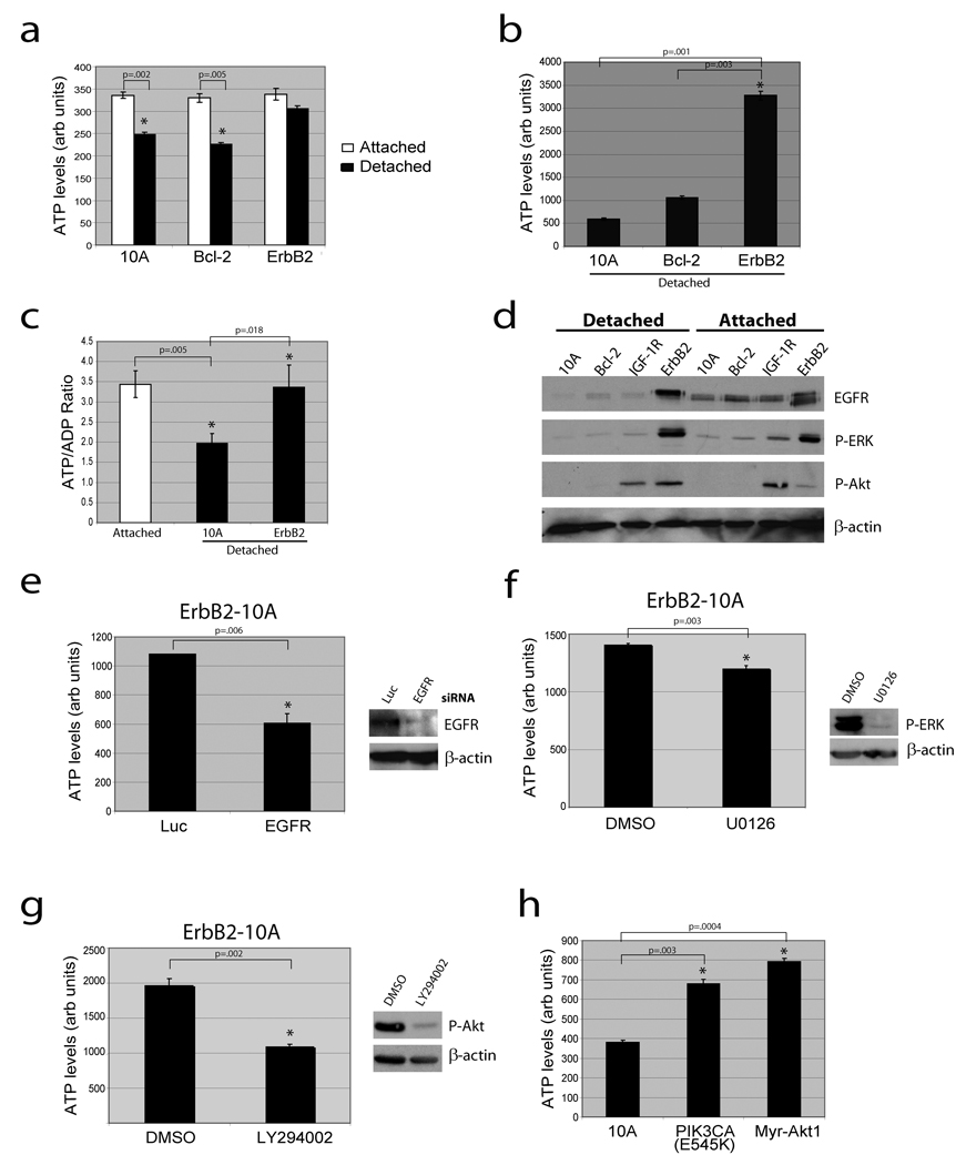
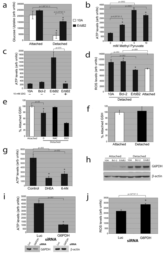
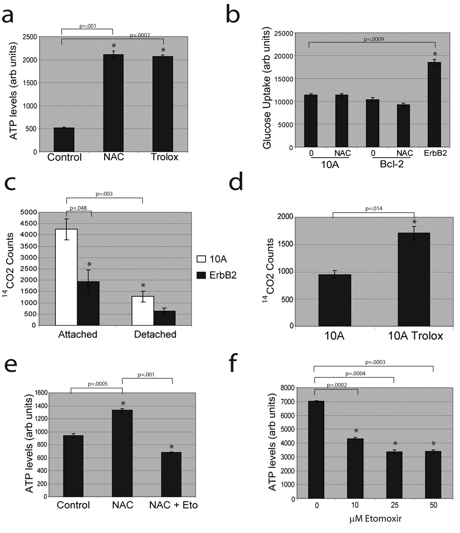
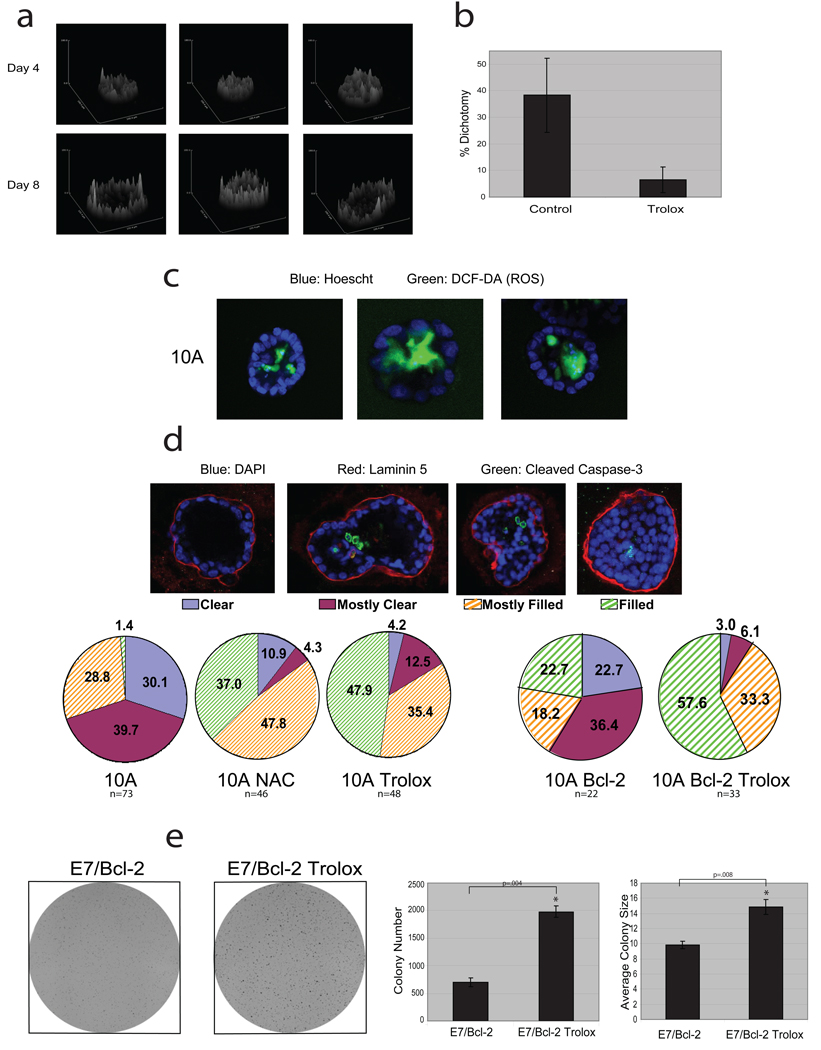
Normal epithelial cells require matrix attachment for survival, and the ability of tumour cells to survive outside their natural extracellular matrix (ECM) niches is dependent on acquisition of anchorage independence. Although apoptosis is the most rapid mechanism for eliminating cells lacking appropriate ECM attachment, recent reports suggest that non-apoptotic death processes prevent survival when apoptosis is inhibited in matrix-deprived cells. Here we demonstrate that detachment of mammary epithelial cells from ECM causes an ATP deficiency owing to the loss of glucose transport. Overexpression of ERBB2 rescues the ATP deficiency by restoring glucose uptake through stabilization of EGFR and phosphatidylinositol-3-OH kinase (PI(3)K) activation, and this rescue is dependent on glucose-stimulated flux through the antioxidant-generating pentose phosphate pathway. Notably, we found that the ATP deficiency could be rescued by antioxidant treatment without rescue of glucose uptake. This rescue was found to be dependent on stimulation of fatty acid oxidation, which is inhibited by detachment-induced reactive oxygen species (ROS). The significance of these findings was supported by evidence of an increase in ROS in matrix-deprived cells in the luminal space of mammary acini, and the discovery that antioxidants facilitate the survival of these cells and enhance anchorage-independent colony formation. These results show both the importance of matrix attachment in regulating metabolic activity and an unanticipated mechanism for cell survival in altered matrix environments by antioxidant restoration of ATP generation.


Figures

Figure 1
Figure 1. Loss of matrix attachment causes reduction in cellular ATP that is rescued by ErbB2 through PI(3)K pathway activation
(a) ATP was measured in the indicated cells 24 hours after plating in adherent or non-adherent (poly-HEMA-coated) plates using the ATPlite assay (a) or the ATP determination kit (b). (c) The ATP/ADP ratio was measured in the indicated cells. (d) The indicated cells were immunoblotted for EGFR, p-ERK, p-Akt, or β-actin. IGF-1R cells were used as a positive control for p-Akt. (e) ErbB2-MCF-10A cells were transfected with siRNAs targeting luciferase (luc) or EGFR and ATP levels were measured using the ATP determination kit. Knockdown was confirmed by immunoblot. (f) and (g) Detached ErbB2-MCF-10A cells were treated with 10 µM UO126 (ERK pathway inhibitor) (f) or 50 µM LY294002 (PI(3)K inhibitor) (g) for 24h and ATP was measured using the ATP determination kit. Inhibition was confirmed by immunoblotting. (h) ATP was measured in detached 10A cells or 10A cells expressing PIK3CA (E545K) or Myr-Akt1 using the ATP determination kit. All error bars represent standard deviation (n=3). A * represents a statistically significant change calculated using a 2 tailed t test.
Figure 2
Figure 2. Matrix detachment causes a reduction in glucose uptake and ErbB2 rescue of this defect is dependent on PPP flux
(a) Glucose uptake was measured in the indicated cells using the Amplex Red assay. (b) ATP was measured using the ATP determination kit after treatment (where indicated) of detached cells with methyl pyruvate. (c) ATP was measured using the ATP determination kit after treatment (where indicated) of detached cells with 2DG. (d) ROS was measured in attached and detached cells using DCF-DA. (e) and (f) Reduced glutathione (GSH) was measured in 10A (e) or ErbB2-MCF-10A (f) cells. Results are graphed as the percent of the attached reduced GSH levels. 1 mM NAC and 1 mM BSO (an inhibitor of glutathione synthesis) were used as a positive and negative control. (g) ATP was measured using the ATP determination kit in detached ErbB2-MCF-10A cells after treatment (where indicated) with vehicle control, 150 µM DHEA, or 150 µM 6-AN. (h) The indicated cell lines were immunoblotted for G6PDH after plating in either normal (attached) or poly-HEMA coated (detached) plates. (i) and (j) ATP (i) and ROS (j) were measured after siRNA mediated knockdown of G6PDH in detached ErbB2-MCF-10A cells. Knockdown was confirmed by immunoblotting. All error bars represent standard deviation (n=3). A * represents a statistically significant change calculated using a 2 tailed t test.
Figure 3
Figure 3. Antioxidants rescue low ATP levels in detached cells by permitting fatty acid oxidation
(a) MCF-10A cells were plated in poly-HEMA coated plates and treated with either control, 1 mM NAC, or 50 µM Trolox and ATP was measured using the ATP determination kit. (b) Detached 10A or Bcl-2-MCF-10A cells were treated with either vehicle (0) or 1 mM NAC and glucose uptake was compared to ErbB2-MCF-10A cells using Amplex Red assay. (c) FAO was measured in parental or ErbB2-MCF-10A cells (either attached or detached). (d) Detached MCF-10A cells were treated with either vehicle (10A) or 50 µM Trolox and FAO was measured. (e) Detached MCF-10A cells were treated with DMSO, 1 mM NAC, and/or 25 µM etomoxir and ATP was measured using the ATP determination kit. (f) Detached ErbB2-MCF-10A cells were treated with the indicated doses of etomoxir and ATP was measured using the ATP determination kit. All error bars represent standard deviation (n=3). A * represents a statistically significant change calculated using a 2 tailed t test.
Figure 4
Figure 4. Analysis of antioxidant effects on acinar morphogenesis and colony formation in soft agar
(a) Native fluorescence of NAD(P)H was assayed by two-photon microscopy in MCF-10A acini. Surface intensity plots of three representative structures from Day 4 or Day 8 are shown. (b) Native fluorescence of NAD(P)H was assayed by two-photon microscopy in MCF-10A acini cultured in the presence or absence of 50 µM Trolox (added daily). At Day 8, structures were blindly scored for dichotomy in fluorescent intensity based on evidence of a reduction in fluorescence intensity in the inner cells (see examples in Supplementary Fig. 14). Error bars represent standard deviation of individual scorings (n=5). (c) ROS was measured in MCF-10A acini at Day 7, by staining with 25 µM DCF-DA (green) and Hoescht (blue). Three representative images are shown and 41% (n=73) of all structures were positive for DCF-DA staining exclusively in the inner cells. NAC or Trolox treatment inhibited the DCF-DA staining in acini (data not shown). (d) Acini were formed using 10A or Bcl-2-MCF-10A cells (10A or 10A Bcl-2) and treated with 1 mM NAC or 50 µM Trolox where indicated. At Day 19 (10A) or Day 33 (Bcl-2), acini were stained for laminin 5, cleaved caspase-3, and with DAPI. Acini were then scored as described. (e) 10A cells expressing E7 and Bcl-2 were plated in soft agar and after 20 days, images were taken after INT-violet staining. Colony number and average colony size were determined using ImageJ. Error bars represent standard deviation (n=3 unless otherwise indicated). A * represents a statistically significant change calculated using a 2 tailed t test.

Comment in

References

    1. Simpson CD, Anyiwe K, Schimmer AD. Anoikis resistance and tumor metastasis. Cancer Lett. 2008 - PubMed
    1. Frisch SM, Screaton RA. Anoikis mechanisms. Curr Opin Cell Biol. 2001;13:555–562. - PubMed
    1. Debnath J, et al. The role of apoptosis in creating and maintaining luminal space within normal and oncogene-expressing mammary acini. Cell. 2002;111:29–40. - PubMed
    1. Mailleux AA, et al. BIM regulates apoptosis during mammary ductal morphogenesis, and its absence reveals alternative cell death mechanisms. Dev Cell. 2007;12:221–234. - PMC - PubMed
    1. Nelson CM, Bissell MJ. Of extracellular matrix, scaffolds, and signaling: tissue architecture regulates development, homeostasis, and cancer. Annu Rev Cell Dev Biol. 2006;22:287–309. - PMC - PubMed

Publication types

MeSH terms